Technology to help doctors find their way around inside knees during surgery
Professor Gustavo Carneiro, Director of Medical Machine Learning, AIML
Story written by Dr Sarah Keenihan, AIML
How’re your knees feeling lately? The knee is a joint that can go through a lot – sometimes just as a part of aging, but also in athletes subjecting their lower limbs to rapid running and turning manoeuvres. Structures inside the knee – such as the meniscus and ligaments – can become torn or rupture, which sometimes calls for surgery to trim or repair.
Along with colleagues, Professor Gustavo Carneiro at the Australian Institute for Machine Learning is developing a new system to help orthopaedic surgeons find their way around inside knees that require surgery. They’ve published their findings as part of the 23rd International Conference on Medical Image Computing and Computer Assisted Intervention.
“Even though it’s a small space, sometimes surgeons find themselves lost inside the knee joint when they’re doing arthroscopic surgery,” said Professor Carneiro.
Arthroscopy is a surgical procedure that avoids the need to open up the whole knee – instead, orthopaedic surgeons insert instruments through button-seized holes to visualize, diagnose, and treat problems inside a joint. At least 70,000 knee arthroscopies are performed every year in Australia.
But arthroscopy can be a difficult procedure, given that the knee joint has only a tiny space (estimated in one study to be less than 4mls in volume) between all its important structures.
“Surgeons are constantly having to work out where their instruments are located in relation to key structures such as bones and ligaments in the knee, and trying to avoid doing any extra damage.”
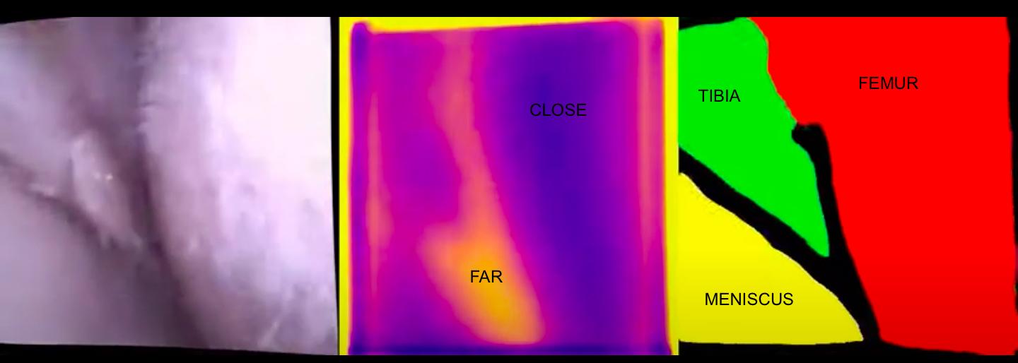
An arthoscopic image of the knee joint is shown at left, with the corresponding images revealing depth information (centre) and anatomical labelling (right).
“Our research aims to provide feedback to the surgeon through live labelling of knee structures in the 3D image that comes from the arthroscopic camera,” said Professor Carneiro.
The machine learning approach being used is called semantic segmentation, where an algorithm is trained to recognise key structures in the knee such as the femur and tibia bones, the anterior cruciate ligament (ACL) and the meniscus (a rubbery structure that acts like a shock absorber in the knee).
“We used more than 8000 images to train our system, with information provided regarding the names of key structures but also depth measurements,” said Professor Carneiro.
“We found this method was much more accurate than a previous, simpler approach to the problem.”
“Our next step is to produce a fully labelled 3D model of the structures in front of the camera, such that surgeons can know the exact position of the arthroscope inside the knee joint,” said Professor Carneiro.
Professor Carneiro is also working on other applications for machine learning in health, including identifying potential cancers in the bowel, picking out abnormalities in chest X-rays, finding cancers that have spread (metastasised) into lymph nodes and detection of endometriosis.
For more information contact Professor Gustavo Carneiro, ARC Future Fellow (2020-2023) at the Australian Institute for Machine Learning, The University of Adelaide.