Preclinical Imaging
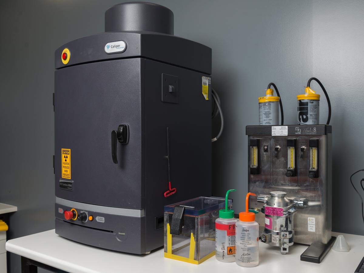
IVIS Lumina XRMS
Please contact Dr Agatha Labrinidis for further information.
Located at Frome Road.
-
Information on the IVIS Lumina XRMS
The IVIS Lumina XRMS Live Animal Biophotonic Imaging System is a real-time in vivo imaging system that offers users the flexibility to image fluorescent and bioluminescent reporters to visualise, monitor and record specific genetic and cellular activity within a living organism.
Bioluminescence Imaging is a high throughput, high-sensitivity, low-noise and non-invasive technique which captures, quantifies and analyses the light emitted by a living organism as the result of a chemical reaction where chemical energy is converted to light energy. This technique can be used to track gene expression and monitor the spread of a disease. It can be applied to drug development.
Bright light on the cancer horizon - a feature story in the Adelaidean.
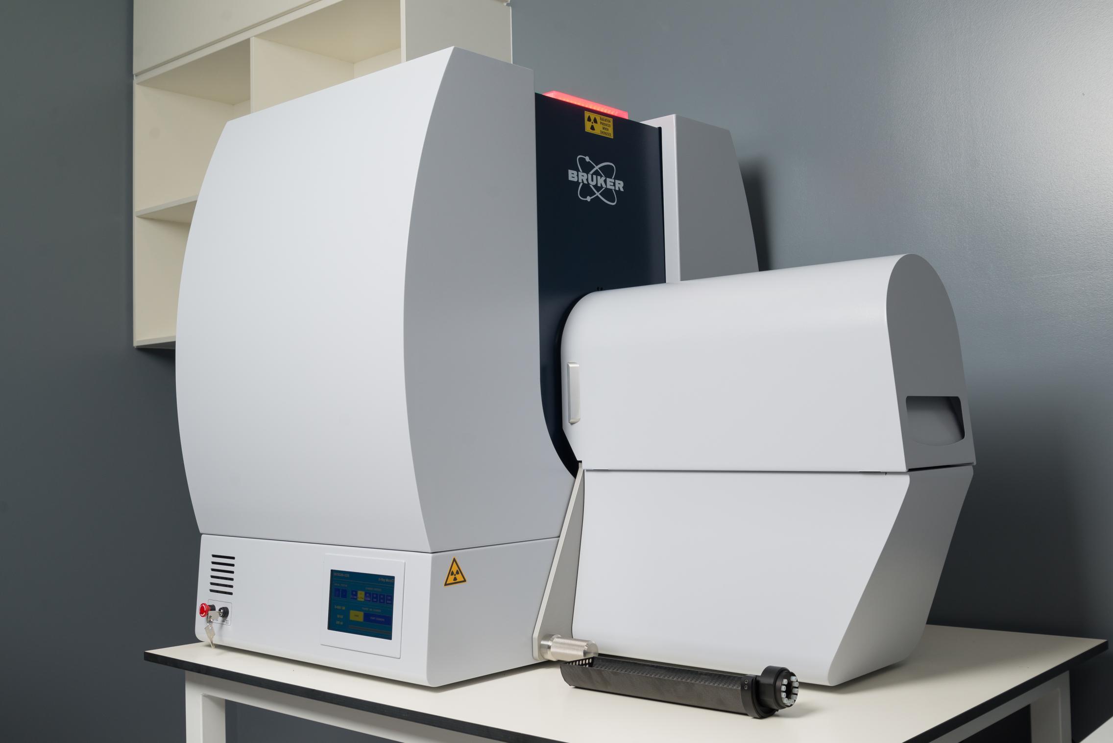
Micro-CT Bruker 1276 Scanner
Please contact Dr Agatha Labrinidis for further information.
Located at Adelaide Health & Medical Sciences Building.
-
Information on the Micro-CT Bruker 1276 Scanner
The 1276 is a high performance, stand-alone, fast, desk-top in vivo micro-CT with continuously variable magnification for scanning small laboratory animals such as mice and rats and also biological samples. It has an unrivaled combination of high resolution, big image size, possibility for round and spiral (helical) scanning and reconstruction, and low dose imaging. The image field of view (up to 80 mm wide and more than 300 mm long) allows full body mouse and rat scanning. The variable magnification allows scanning bone and tissue samples with high spatial resolution down to 2.8µm pixel size. Variable X-Ray energy combined with a range of filters ensures optimal image quality for diverse research applications from lung tissue to bone with metal implants. Further, the 1276 in vivo micro-CT administers low radiation dose to the animals allowing multiple scans in longitudinal pre-clinical studies without the risk of unwanted radiation-induced side effects. The system can perform scanning with continuous gantry rotation and in step-and-shoot mode with fastest scanning cycle 3.9 sec.
The 1276 allows reconstructing non-invasively any cross section(s) through the animal body with possibilities to convert reconstructed datasets into realistic 3D-image and calculate internal morphological parameters including specific bone structural parameters.
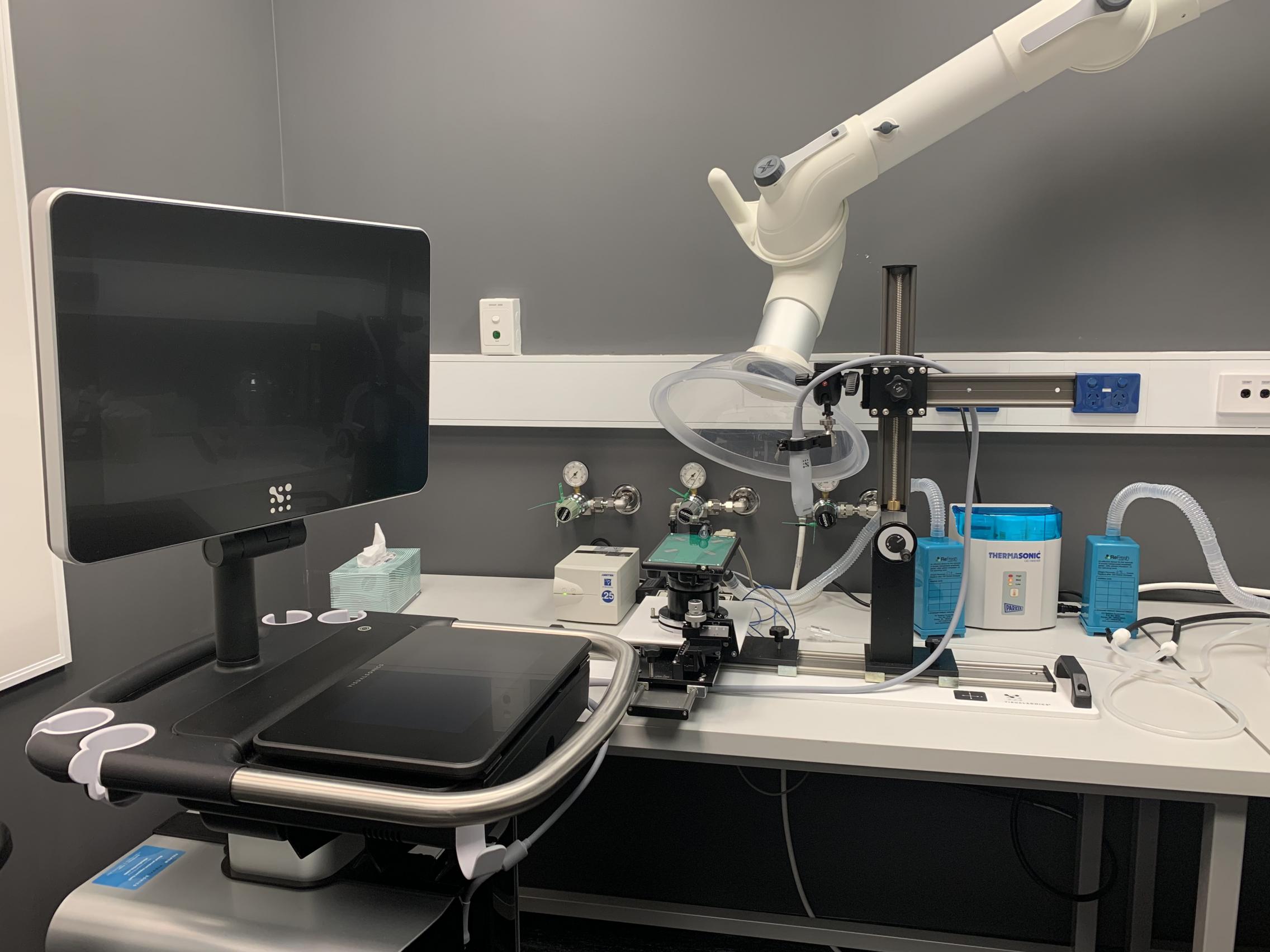
Ultrasound Vevo3100 Preclinical Imaging
Please contact Dr Agatha Labrinidis for further information.
Located at Adelaide Health & Medical Sciences Building.
-
Information on the Ultrasound Vevo3100 Preclinical Imaging
Ultrasound is sound waves with frequencies which are higher than those audible to humans (>20,000 Hz). Ultrasonic images, also known as sonograms, are made by sending pulses of ultrasound into tissue using a probe.
The sound echoes off the tissue; with different tissues reflecting varying degrees of sound. These echoes are recorded and displayed as an image to the operator.
It is used to see internal body structures such as tendons, muscles, joints, blood vessels, and internal organs. Its aim is often to find a source of a disease or to exclude any pathology.
Applications include:
- Oncology- Tumor detection 2D, 3D, Vascularity and perfusion, Tumor model characterization, response to therapy
- Abdominal- Kidney Function, Liver Fibrosis, Reproductive
Cardiovascular- Cardiac Function in 2D, 3D and 4D, Hemodynamics, Myocardinal and vascular strain, Cardiotoxicity - Developmental- Placental structure and function, pregnancy screening, Image-guided embryo injections
- Heamatology- Thrombosis, Hemodynamics, blood velocity
- Molecular Biology- Microdistribution of biomarkers
- Nephrology- Kidney Function, Evaluate renal microcirculatory flow, Early detection of cysts, tumors, obstructions, etc., Renal biopsy and/or injection
- Neurobiology- Total Hgb and blood velocity, Glioma research, Stroke assessment,
- Toxicology
- Gene Deliver
- Image-Guided Injections
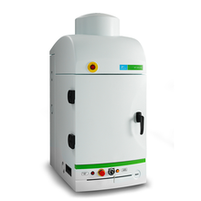
IVIS Lumina X5 Preclinical Imaging
Please contact Dr Agatha Labrinidis for further information.
Located at Adelaide Health & Medical Sciences Building.
-
Information on the IVIS Lumina X5 Preclinical Imaging
The IVIS Lumina X5 is a real-time in vivo imaging system that offers users the flexibility to image fluorescent and bioluminescent reporters to visualise, monitor and record specific genetic and cellular activity within a living organism. Bioluminescence Imaging is a high throughput, high-sensitivity, low-noise and non-invasive technique which captures, quantifies and analyses the light emitted by a living organism as the result of a chemical reaction where chemical energy is converted to light energy. This technique can be used to track gene expression, monitor the spread of a disease and assist in drug development. In addition it includes a high resolution X-ray, allowing the overlay of signals over the skeleton which may have underlying anatomical and structural changes.
For more information please see product note attached. For training and access please contact Agatha Labrinidis.