Microsporum
The genus Microsporum is now restricted to just three species: M. audouinii, M. canis and M. ferrugineum.
The remaining geophilic and zoophilic species, previously considered Microsporum species, have been transferred to the genera Lophophyton, Nannizzia and Paraphyton (de Hoog et al. 2017).
-
Laboratory Identification
Microsporum species may form both macro- and microconidia, although they are not always present. Cultures are mostly granular to cottony, yellowish to brownish, with a cream-coloured or brown colony reverse. Macroconidia are hyaline, multiseptate, with thick rough cell walls, and are clavate, fusiform or spindle-shaped. Microconidia are single-celled, hyaline, smooth-walled, and are predominantly clavate in shape.
Note:
Strains of M. canis often do not produce macroconidia and/or microconidia on primary isolation media and subcultures onto polished rice grains or lactritmel agar are recommended to stimulate sporulation. These non-sporulating strains of M. canis are often erroneously identified as M. audouinii and it is surprising just how many laboratories have difficulty in differentiating M. canis and M. audouinii.Molecular Identification:
ITS sequencing is recommended (Gräser et al. 1998, 2000, Brillowska-Dabrowska et al. 2013).References:
Rebell and Taplin (1970), Rippon (1988), McGinnis (1980), Domsch et al. (1980), Ajello (1977), Weitzman et al.(1986), Mackenzie et al. (1986), Kane et al. (1997), de Hoog et al. (2000, 2015), Gräser et al. (1999a, 2008). Cafarchia et al. (2013).
Species descriptions
-
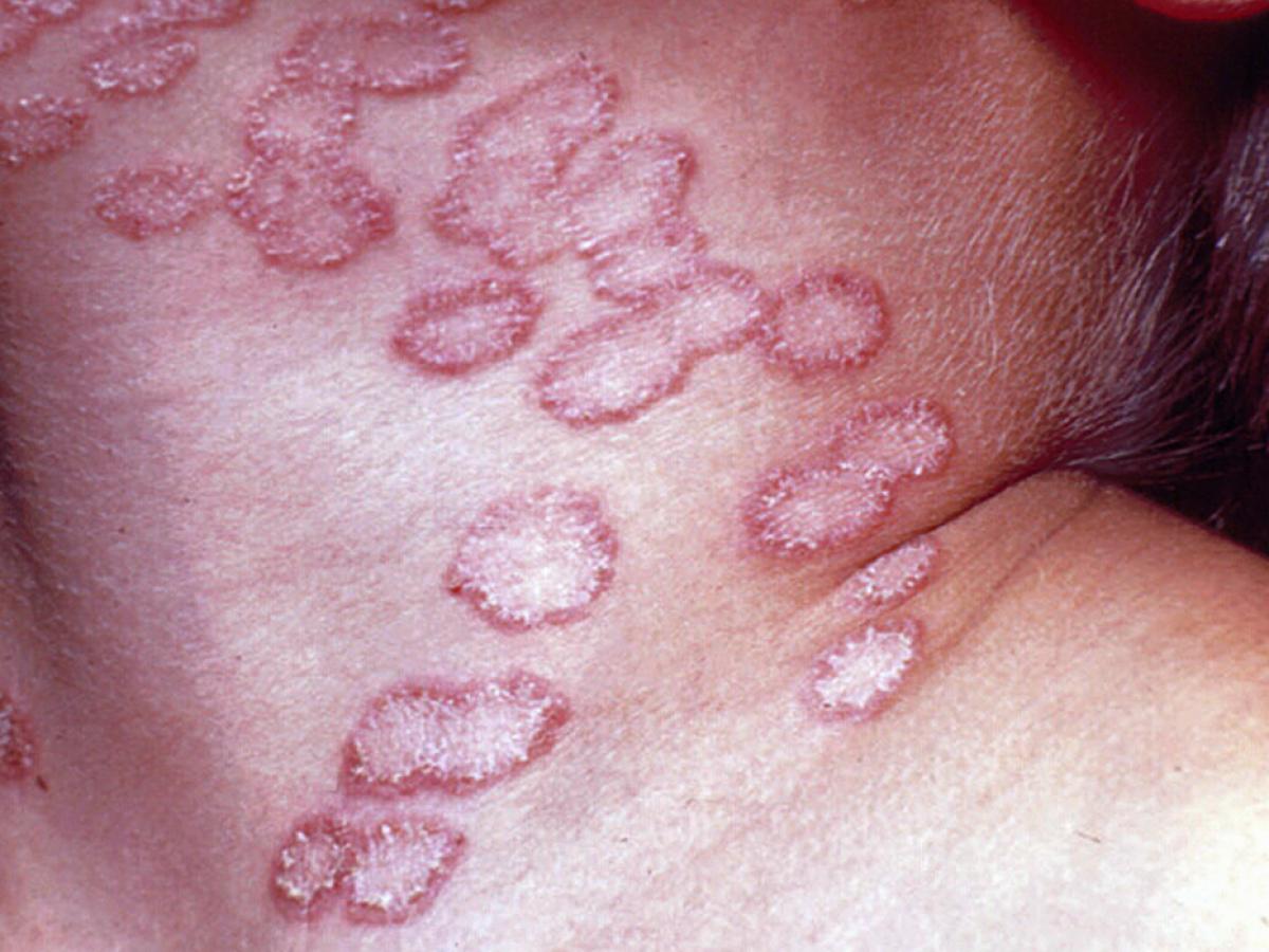
Microsporum audouinii
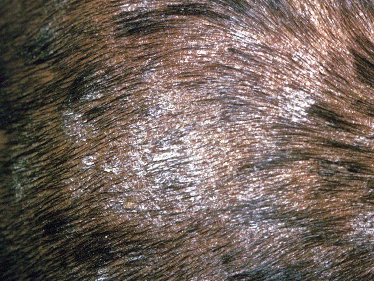
Tinea capitis caused by Microsporum audouinii.
Microsporum audouinii is an anthropophilic fungus causing non-inflammatory infections of the scalp and skin, especially in children. Once the cause of epidemics of tinea capitis in Europe and North America, it is now less common. Invaded hairs show an ectothrix infection and usually fluoresce a bright greenish-yellow under Wood’s ultra-violet light. Only rarely found in Australasia, most reports are in fact misidentified non-sporulating strains of M. canis.
RG-2 organism.
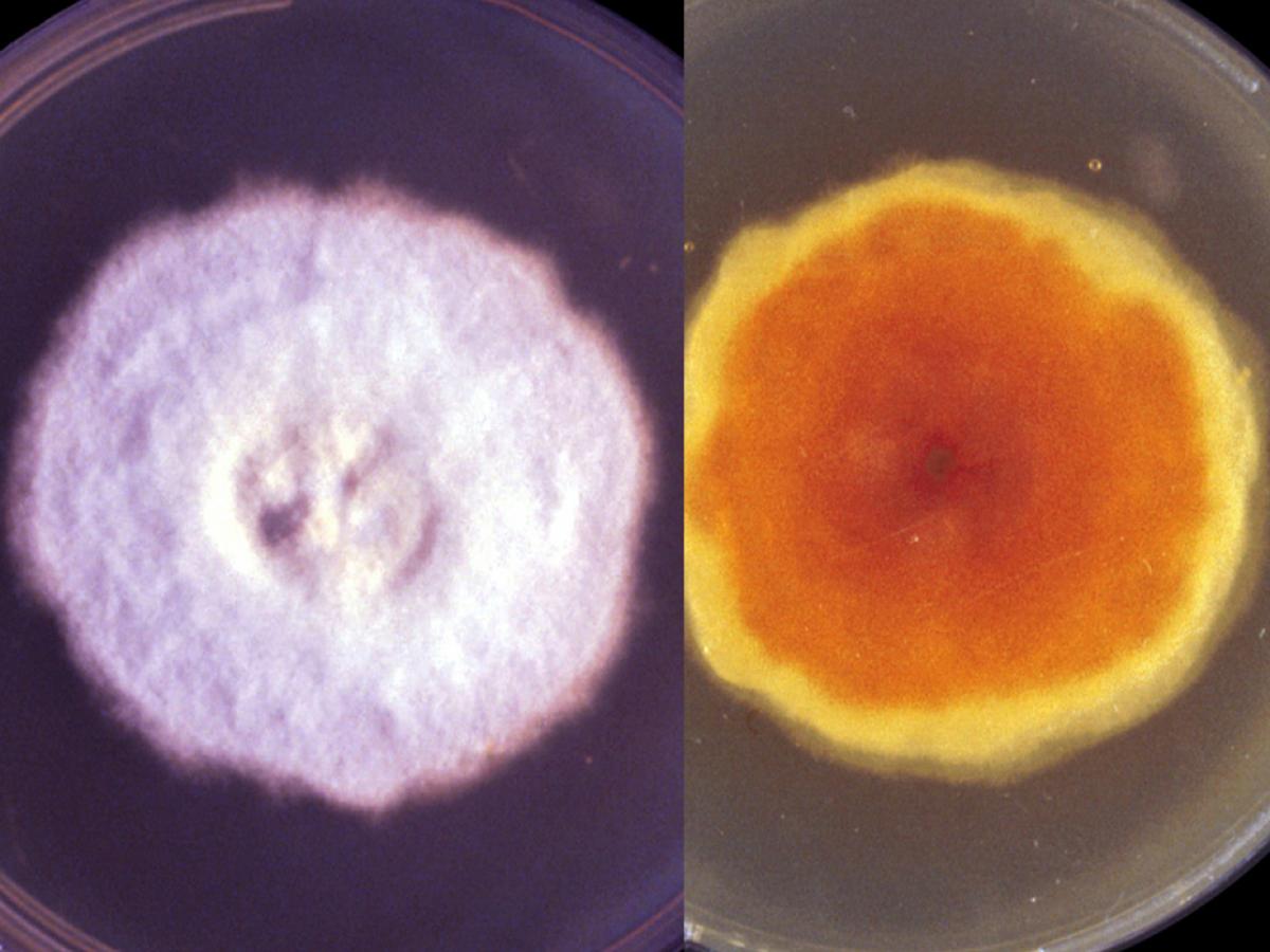
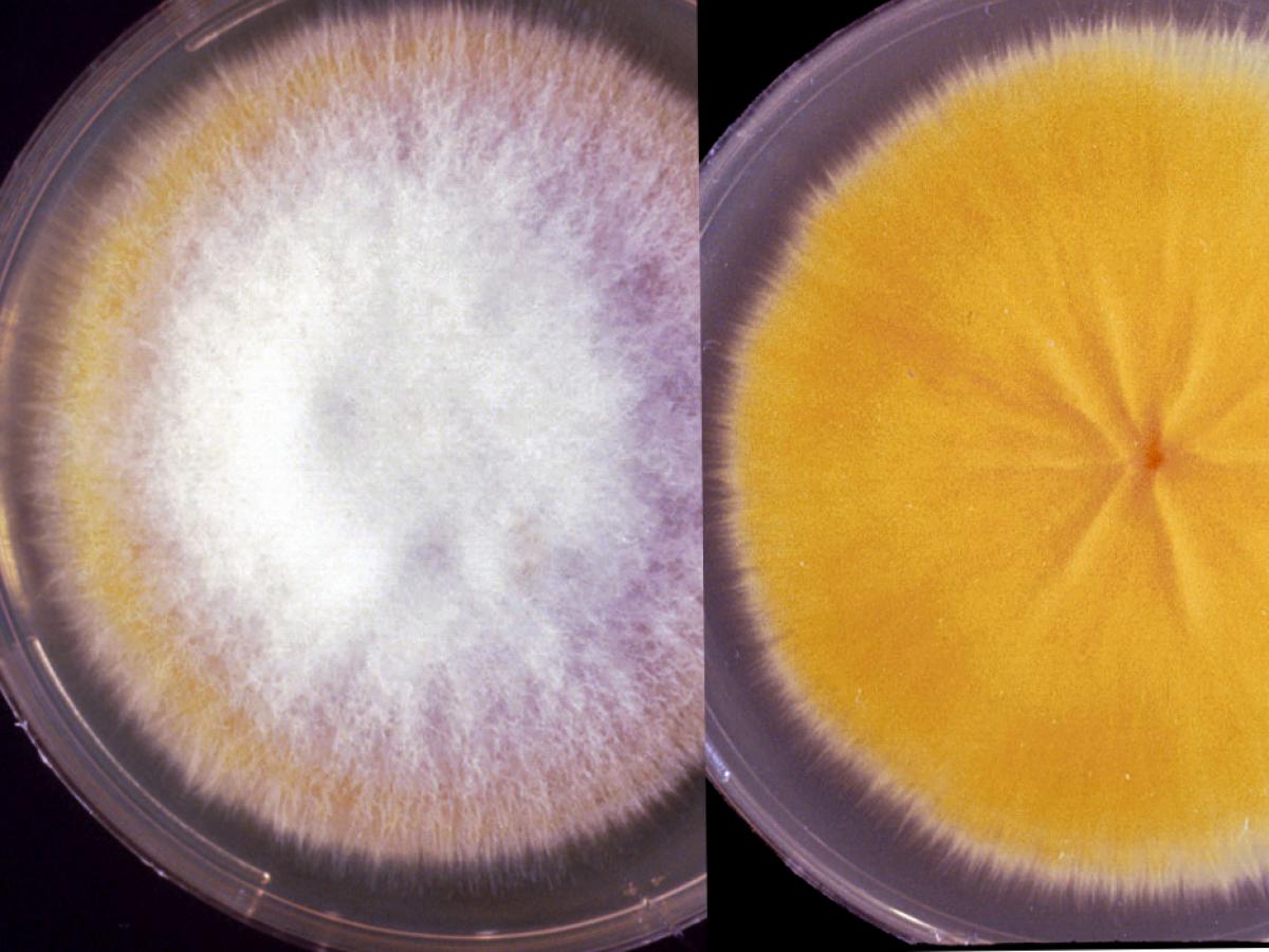
Culture of Microsporum audouinii..
Morphological description:
Colonies are flat, spreading, greyish-white to light tan-white in colour, and have a dense suede-like to downy surface, suggestive of mouse fur in texture. Reverse can be yellow-brown to reddish-brown in colour. Some strains may show no reverse pigment. Macroconidia and microconidia are rarely produced, most cultures are sterile or produce only occasional thick-walled terminal or intercalary chlamydospores. When present, macroconidia may resemble those of M. canis but are usually longer, smoother and more irregularly fusiform in shape; microconidia, when present, are pyriform to clavate in shape and are similar to those seen in other species of Microsporum, Lophophyton and Nannizzia. Pectinate (comb-like) hyphae and racquet hyphae (a series of hyphal segments swollen at one end) may also be present.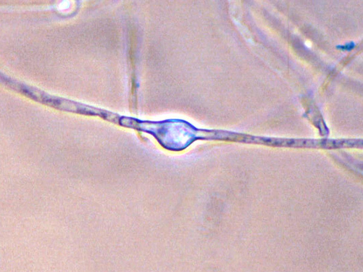
Chlamydoconidium of M. audouinii.
Confirmatory tests:
Growth on rice grains:
Very poor or absent, usually being visible only as a brown discolouration. This is one of the features which distinguish M. audouinii from M. canis.Reverse pigment on potato dextrose agar:
Salmon to pinkish-brown (M. canis is bright yellow).BCP milk solids glucose agar:
Both M. canis and M. audounii demonstrate profuse growth, but only M. audouinii shows a rapid pH change to alkaline (purple colour).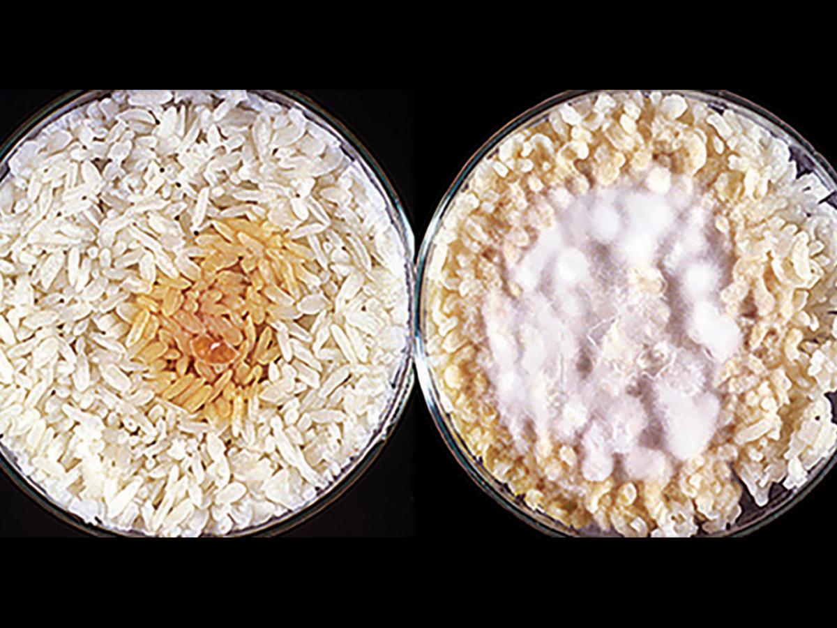
(left) Microsporum audouinii showing poor growth on rice grains, usually being visible only as a brown discolouration and (right) Microsporum canis on rice grains showing good growth, yellow pigmentation and sporulation.
Vitamin free agar (Trichophyton agar No.1):
Good growth indicating no special nutritional requirements. Cultures are flat, white, suede-like to downy, with a yellow-brown reverse. Note: Growth of some strains of M. audouinii is enhanced by the presence of thiamine (Trichophyton agar No.4).Hair perforation test:
Negative after 28 days.Key features:
Absence of conidia, poor or no growth on polished rice grains, inability to perforate hair in vitro, and culture characteristics. -
Microsporum canis
Dermatophytosis (ringworm) caused by Microsporum canis.
Microsporum canis is a zoophilic dermatophyte of worldwide distribution and is a frequent cause of ringworm in humans, especially children. Invades hair, skin and rarely nails. Cats and dogs are the main sources of infection. Invaded hairs show an ectothrix infection and fluoresce a bright greenish-yellow under Wood’s ultra-violet light.
RG-2 organism.
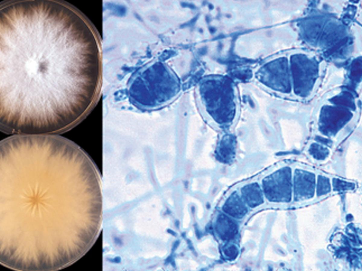
Culture of Microsporum canis.
Morphological description:
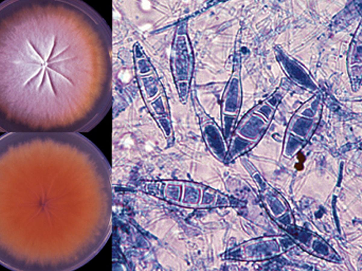
Colonies are flat, spreading, white to cream-coloured, with a dense cottony surface which may show some radial grooves. Colonies usually have a bright golden yellow to brownish yellow reverse pigment, but non-pigmented strains may also occur. Macroconidia are typically spindle-shaped with 5-15 cells, verrucose, thick-walled and often have a terminal knob, 35-110 x 12-25 µm. A few pyriform to clavate microconidia are also present. Macroconidia and/or microconidia are often not produced on primary isolation media and it is recommended that subcultures be made onto lactritmel agar and/or boiled polished rice grains to stimulate sporulation.
Macroconidia of Microsporum canis are typically spindle-shaped with 5-15 cells, verrucose, thick-walled and often have a terminal knob. A few pyriform to clavate microconidia are also present.
Growth on rice grains:
Good growth of white aerial mycelium with production of yellow pigment. Microscopy reveals numerous macroconidia and microconidia similar to those described above. Note: Macroconidia and/or microconidia of M. canis are often not produced on primary isolation media and it is recommended that sub-cultures be made onto boiled polished rice grains to stimulate sporulation.Reverse pigment on potato dextrose agar:
Bright yellow (both M. audouinii and M. canis var. equinum are salmon to pinkish-brown).Vitamin free agar (Trichophyton agar No.1):
Good growth indicating no special nutritional requirements. Cultures are flat, white, suede-like to downy, with a yellow to pale yellow-brown reverse.Hair perforation test:
Positive at 14 days.Key features:
Distinctive macroconidia and culture characteristics. Abundant growth and sporulation on polished rice grains and in vitro perforation of hair.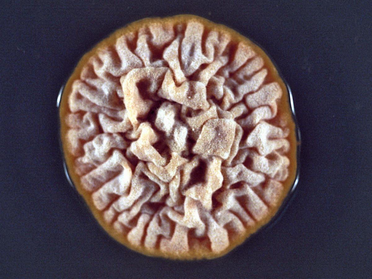
Microsporum canis dysgonic strain.
Microsporum canis dysgonic strains are rare but may also occur. These dysgonic strains typically have a heaped and folded, yellow-brown thallus and macroconidia are usually absent. However, typical colonies and macroconidia of M. canis are usually produced by this variant when subcultured onto polished rice grains. Note: The dysgonic type colony of M. canis is similar to that of Microsporum ferrugineum.
Microsporum canis var. distortum.
Supplementary description for Microsporum canis var. distortum, a dysgonic variant of M. canis with distinctive distorted macroconidia. Abundant growth and sporulation on rice grains.
Microsporum canis var. distortum is a zoophilic fungus known to cause infections in cats, dogs and other animals. It is a rare cause of tinea capitis in New Zealand, Australia and North America. Clinical disease is similar to M. canis. Invaded hairs show an ectothrix infection and fluoresce a bright greenish-yellow under Wood’s ultra-violet light.
Microsporum canis var. equinum.
Supplementary description for Microsporum canis var. equinum which is now considered to be a genotypic synonym of Microsporum canis (de Hoog et al. 2000). This variant of M. canis is a rare cause of ringworm of horses. Invaded hairs show an ectothrix infection and fluoresce a bright greenish-yellow under Wood’s ultra-violet light. Rarely infects man or other animal species. Reported from Australia, Europe and North America.
-
Microsporum ferrugineum
Microsporum ferrugineum is an anthropophilic fungus causing epidemic juvenile tinea capitis in humans. The clinical features are similar to those of infections caused by M. audouinii. Invaded hairs show an ectothrix infection and fluoresce a greenish-yellow under Wood’s ultra-violet light. Reported from Asia (including China and Japan), Russia, Eastern Europe and Africa.
RG-2 organism.

Culture and Bamboo hyphae of M. ferrugineum.
Morphological description:
Colonies are slow growing, forming a waxy, glabrous, convoluted thallus with a cream to buff-coloured surface and no reverse pigment. Note: Surface pigmentation may vary from cream to yellow to deep red and a flatter white form sometimes occurs. Cultures rapidly become downy and pleomorphic. Microscopic morphology is negative, microconidia or macroconidia are not produced. However, irregular branching hyphae with prominent cross walls (“bamboo hyphae”) and chlamydospores are seen. “Bamboo hyphae” are a characteristic of this species.Key features:
Clinical history, culture characteristics and distinctive “bamboo hyphae”.