Nannizzia
Based on a recent multilocus phylogenetic study the taxonomy of the dermatophytes has been reviewed. The genus Nannizzia now consists of 9 species (de Hoog et al. 2017).
Colonies are mostly cottony to powdery, whitish to brown, with a cream-coloured, brown or red. Macroconidia are 2- or multicelled, hyaline, cylindrical, or clavate to cigar-shaped and smooth-walled. Microconidia are hyaline, 1-celled, ovoidal, pyriform to clavate and smooth-walled.
Note: N. fulva, N. gypsea, N. nana and N. persicolor were previously included in the genus Microsporum.
Species descriptions
-
Nannizzia fulva
Synonymy:
Microsporum fulvumNannizzia fulva is a geophilic fungus of worldwide distribution which may cause occasional infections in humans and animals. Clinical disease is similar to N. gypsea but less common. Invaded hairs show a sparse ectothrix infection but do not fluoresce under Wood’s ultra-violet light.
RG-1 organism.
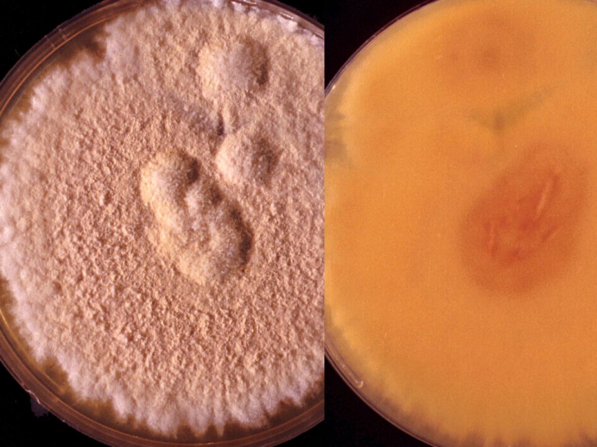
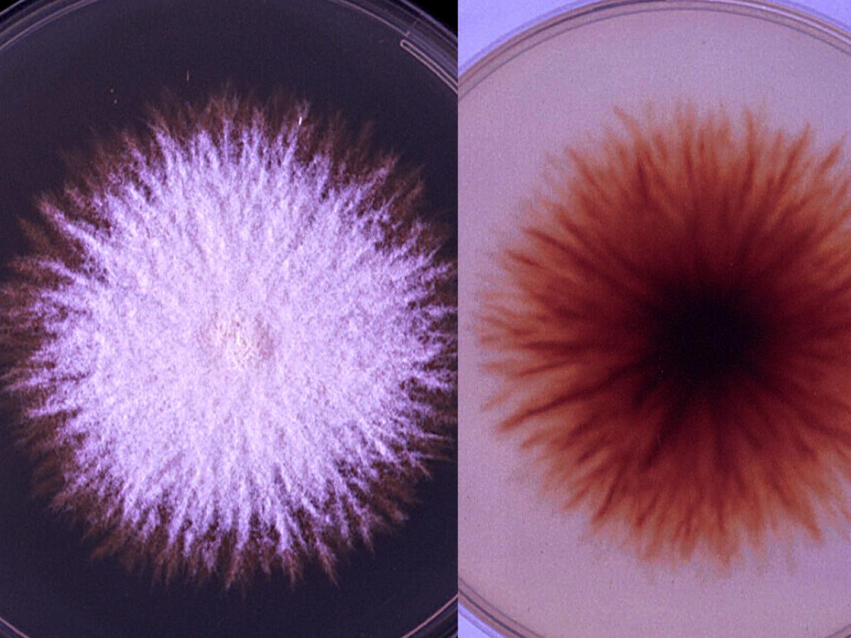
Nannizzia fulva culture.
Morphological description:
Colonies are fast growing, flat, suede-like, tawny-buff to pinkish-buff in colour and frequently have a fluffy white advancing edge. A dark red under surface is occasionally seen, otherwise it is colourless to yellow brown. Abundant thin-walled, elongate, ellipsoidal macroconidia are formed which closely resemble those of N. gypsea, except they are longer and more bullet-shaped (clavate) with three to six septa. Numerous spiral hyphae, which are often branched are seen. Numerous pyriform to clavate microconidia are also produced but these are not diagnostic.
Nannizzia fulva macroconidia.
Vitamin free agar (Trichophyton agar No.1):
Good growth indicating no special nutritional requirements. Cultures are flat, white, suede-like to downy, with a yellow-brown reverse. Note: Growth of some strains of M. audouinii is enhanced by the presence of thiamine (Trichophyton agar No.4).Hair perforation test:
Negative after 28 days.Key features:
Absence of conidia, poor or no growth on polished rice grains, inability to perforate hair in vitro, and culture characteristics. -
Nannizzia gypsea
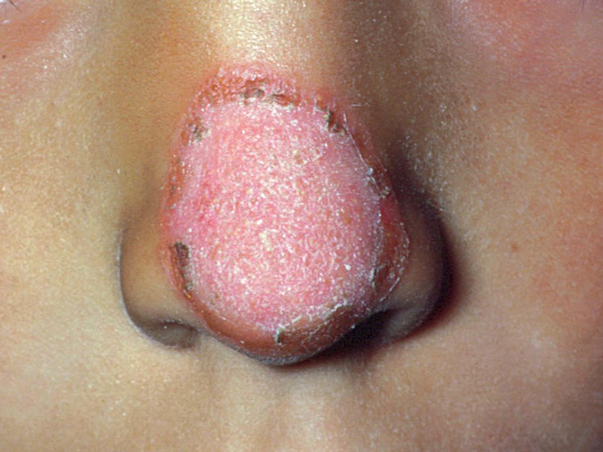
Dermatophytosis (ringworm) caused by Nannizzia gypsea.
Nannizzia gypsea is a geophilic fungus with a worldwide distribution which may cause infections in animals and humans, particularly children and rural workers during warm humid weather. Usually produces a single inflammatory skin or scalp lesion. Invaded hairs show an ectothrix infection but do not fluoresce under Wood’s ultra-violet light.
RG-1 organism.
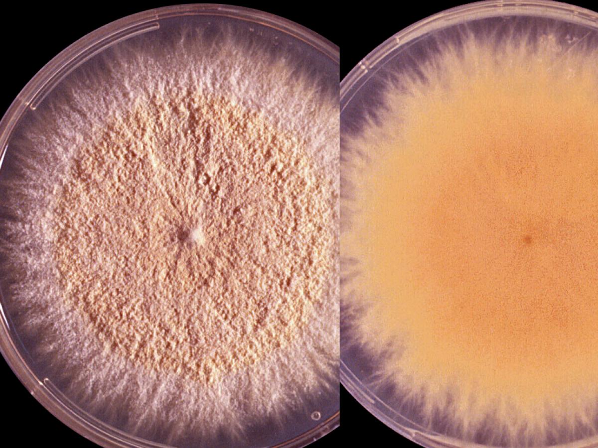
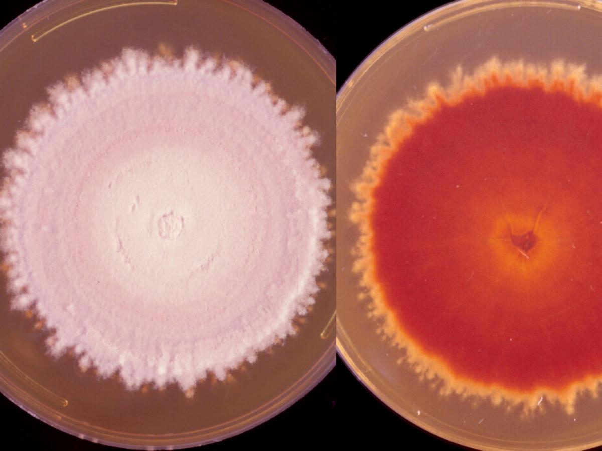
Culture of Nannizzia gypsea.
Morphological description:
Colonies are usually flat, spreading, suede-like to granular, with a deep cream to tawny-buff to pale cinnamon-coloured surface. Many cultures develop a central white downy umbo (dome) or a fluffy white tuft of mycelium and some also have a narrow white peripheral border. A yellow-brown pigment, often with a central darker brown spot, is usually produced on the reverse, however a reddish-brown reverse pigment may be present in some strains. Cultures produce abundant, symmetrical, ellipsoidal, thin-walled, verrucose, four to six-celled macroconidia. The terminal or distal ends of most macroconidia are slightly rounded, while the proximal ends (point of attachment to hyphae) are truncate. Numerous clavate-shaped microconidia are also present, but these are not diagnostic.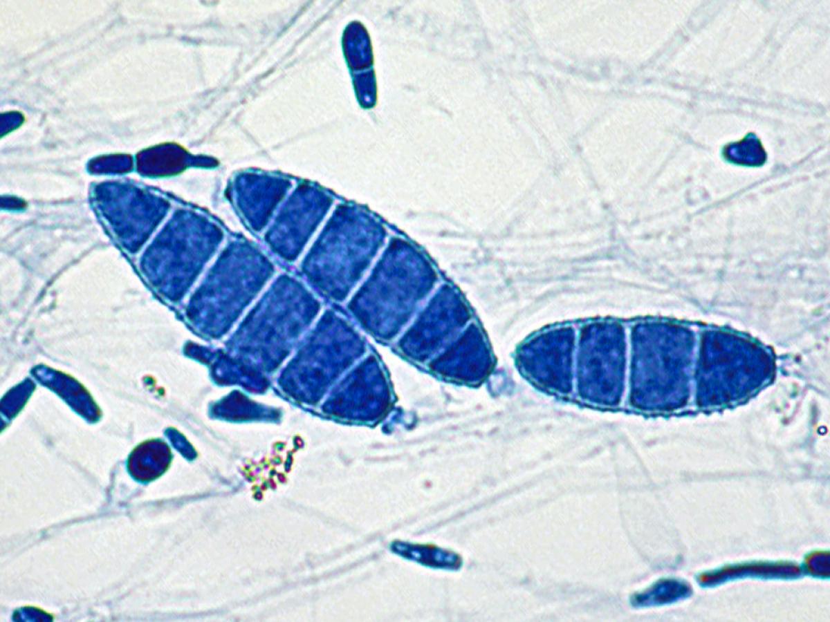
Macroconidia of Nannizzia gypsea are typically symmetrical, ellipsoidal, thin-walled, verrucose, four to six-celled macroconidia. Numerous pyriform to clavate microconidia are also present.
Key features:
Distinctive macroconidia and culture characteristics.Molecular identification:
ITS sequencing is recommended, especially for the separation of N. gypsea and N. incurvata which are morphologically similar.References:
Rebell and Taplin (1970), Rippon (1988), Gräser et al. (2008), Cafarchia et al. (2013), de Hoog et al. (2015, 2016). -
Nannizzia nana
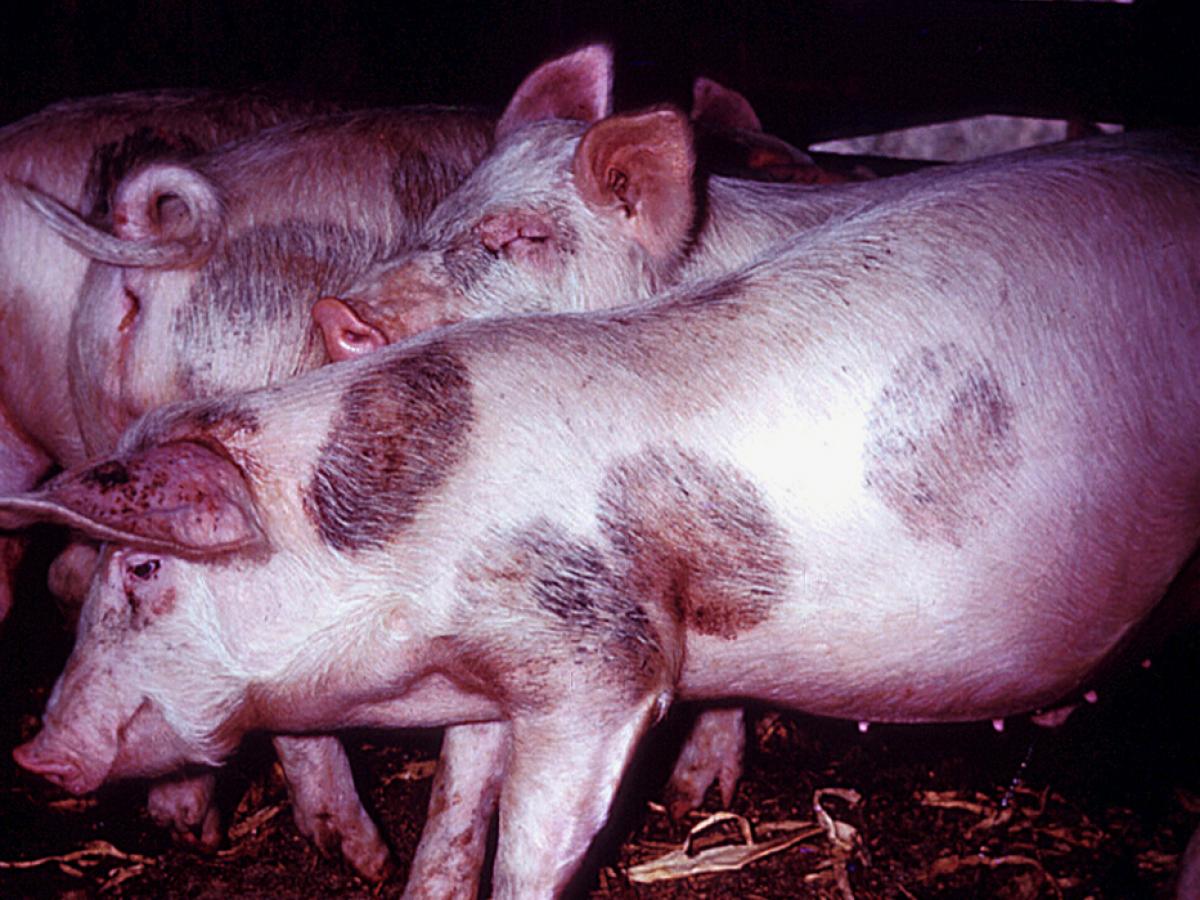
Chronic non-inflammatory lesions in pigs caused by N. nana.
Synonymy:
Microsporum nanumNannizzia nana is a geophilic and zoophilic fungus frequently causing chronic non-inflammatory lesions in pigs and a rare cause of tinea in humans. Also present in soil of pig-yards. Infections in man are usually contracted directly from pigs or fomites. Invaded hairs may show a sparse ectothrix or endothrix infection but do not fluoresce under Wood’s ultra-violet light. The geographical distribution is worldwide.
RG-2 organism.
Culture of Nannizzia nana.
Morphological description:
Colonies are flat, cream to buff in colour with a suede-like to powdery surface texture. Young colonies have a brownish-orange pigment which deepens into a dark reddish-brown with age. Cultures produce numerous small ovoid to pyriform macroconidia with one to three (mostly two) cells, with relatively thin, finely echinulate (rough) walls, and broad truncate bases. Many macroconidia are borne on conidiophores (stalks) which do not stain readily. Occasional clavate microconidia are present, which distinguishes N. nana from some species of Chrysosporium.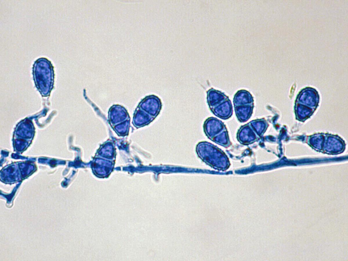
Macroconidia of Nannizzia nana.
Key features:
Distinctive macroconidia and culture characteristics.Molecular identification:
ITS sequencing is recommended.References:
Rebell and Taplin (1970), Rippon (1988), Gräser et al. (2008), Cafarchia et al. (2013), de Hoog et al. (2015, 2016). -
Nannizzia persicolor
Synonymy:
Microsporum persicolorNannizzia persicolor is a zoophilic fungus often occurring as a saprophyte on voles and bats. A rare cause of tinea corporis in humans. Not known to invade hair in vivo, but produces hair perforations in vitro. Distribution: Africa, Australia, Europe and North America.
RG-2 organism.
Culture of Nannizzia persicolor.
Morphological Description:
Colonies are generally flat, white to pinkish in colour, with a suede-like to granular texture and peripheral fringe. Reverse pigmentation is orange to red. Macroconidia are thin-walled, cigar-shaped, four to seven-celled, 40-60 x 6-8 µm but are only rarely produced. Microconidia are abundant, spherical to pyriform.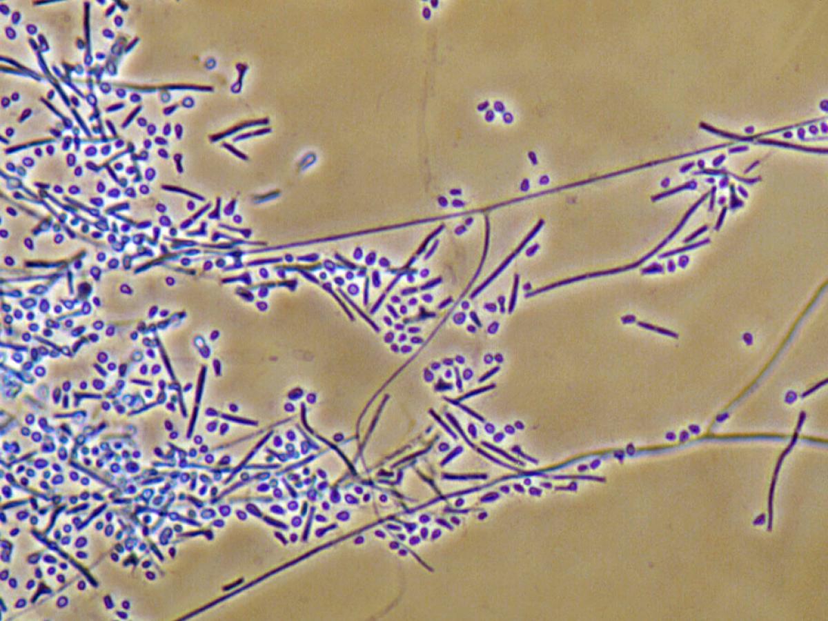
Microconidia of Nannizzia persicolor.
Key Features:
Microscopic morphology and culture characteristics.Molecular Identification:
ITS sequencing is recommended.References:
Rebell and Taplin (1970), Rippon (1988), Gräser et al. (2008), Cafarchia et al. (2013), de Hoog et al. (2015, 2016).