Acrophialophora fusispora
The genus Acrophialophora contains 16 species that are most commonly associated with soil, especially from India (Zhang et al. 2015a).
Two species have been reported as human pathogens, A. fusispora and A. levis (Sandoval-Denis et al. 2015). A. levis conidia lack spiral bands and the phialides have verruculose walls, ITS sequencing is recommended for species identification.
RG-1 organism.
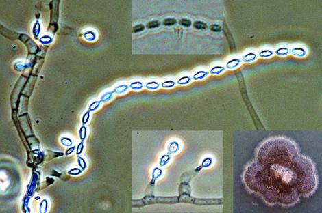
Culture, phialides and conidia with striations of A. fusispora.
Morphological description:
Colonies fast growing, greyish-brown with a black reverse. Conidiophores arising singly, terminally and laterally from the hyphae, erect, straight or slightly flexuose, tapering towards the apex, pale brown, rough-walled, up to 15 μm long, 2-5 μm wide, with whorls of phialides on the upper part. Phialides flask-shaped with a swollen base and a long, narrow neck, hyaline, smooth-walled or echinulate, 9-15 x 3-4.5 μm in the broadest part. Conidia in long chains, limoniform, one-celled, pale brown 5-12 x 3-6 μm, smooth to finely echinulate with indistinct spiral bands. Temperature: optimum 40C; maximum 50C.
Key features:
Hyphomycete with flask-shaped phialides producing long chains of one-celled, limoniform, pale brown conidia, with indistinct spiral bands. Note: in A. levis the conidia lack spiral bands and the phialides have verruculose walls.
Molecular identification:
D1/D2, ITS, 18S and β-tubulin sequences have been reported by Sandoval-Denis et al. (2015) and Zhang et al. (2015a).
| Antifungal | Range | MIC90 | Antifungal | Range | MIC90 |
|---|---|---|---|---|---|
| AmB | 1-32 | 16 | VORI | 0.06-0.5 | 0.25 |
| ITRA | 0.125-4 | 1 | POSA | 0.25-1 | 1 |
References: Domsch et al. (2007), de Hoog et al. (2000, 2015), Al-Mohsen et al. (2000), Guarro et al. (2007), Sandoval-Denis et al. (2015), Zhang et al. (2015a).