Cladophialophora
The genus Cladophialophora is characterised by: (1) the absence of conidiophores, “shield cells,” or prominent hila (attachment points); (2) the ability to grow on media containing cycloheximide; and (3) having dry, non-fragile chains of conidia (Revankar and Sutton 2010).
It has recently been re-evaluated by multilocus sequencing and currently contains seven species associated with human infection (Badali et al. 2008).
Cladophialophora bantiana is the causative agent of numerous cases of cerebral phaeohyphomycosis many of which occur in immunocompetent individuals and most of which are fatal (Chakrabarti et al. 2016). C. carrionii and the recently described C. samoensis are agents of chromoblastomycosis. Less common species occasionally implicated in deep and superficial mycoses include, C. arxii, C. boppii, C. devriesii, C. emmonsii, C. modesta and C. saturnica. C. yegresii is a closely related environmental sister species to C. carrionii (Revankar and Sutton 2010, de Hoog et al. 1995, 2015).
Species descriptions
-
Cladophialophora bantiana
Synonymy:
Xylohypha bantiana, Cladosporium bantianum.Cladophialophora bantiana has been isolated from soil and is a recognised agent of cerebral phaeohyphomycosis. The fungus is neurotropic and may cause brain abscess in both normal and immunosuppressed patients and is usually fatal. The fungus is likely introduced via inhalation and direct transfer to the brain via the paranasal sinuses, or traumatic head injury.
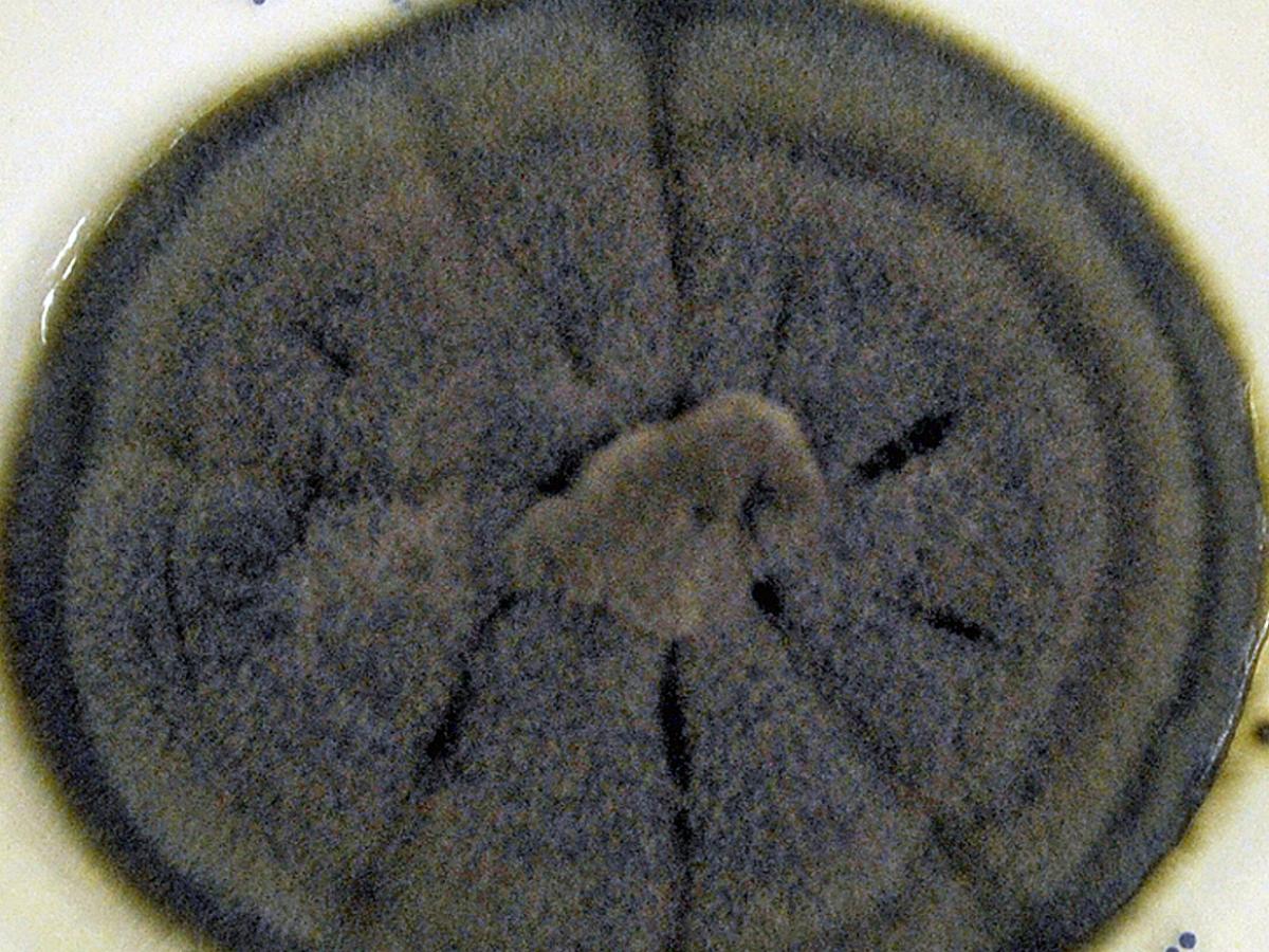
Cladophialophora bantiana culture.
WARNING: RG-3 organism.
Cultures of C. bantiana represent a potential biohazard to laboratory personnel and must be handled with extreme caution in Class II Biological Safety Cabinet (BSCII).Morphological description:
Colonies are moderately fast growing, olivaceous-grey, suede-like to floccose and grow at temperatures up to 42-40C. Conidia are formed in long, sparsely branched, flexuose, acropetal chains from undifferentiated conidiophores. Conidia are one-celled (very occasionally two-celled), pale brown, smooth-walled, ellipsoid to oblong-ellipsoid and are 2-3 x 4-7 µm in size.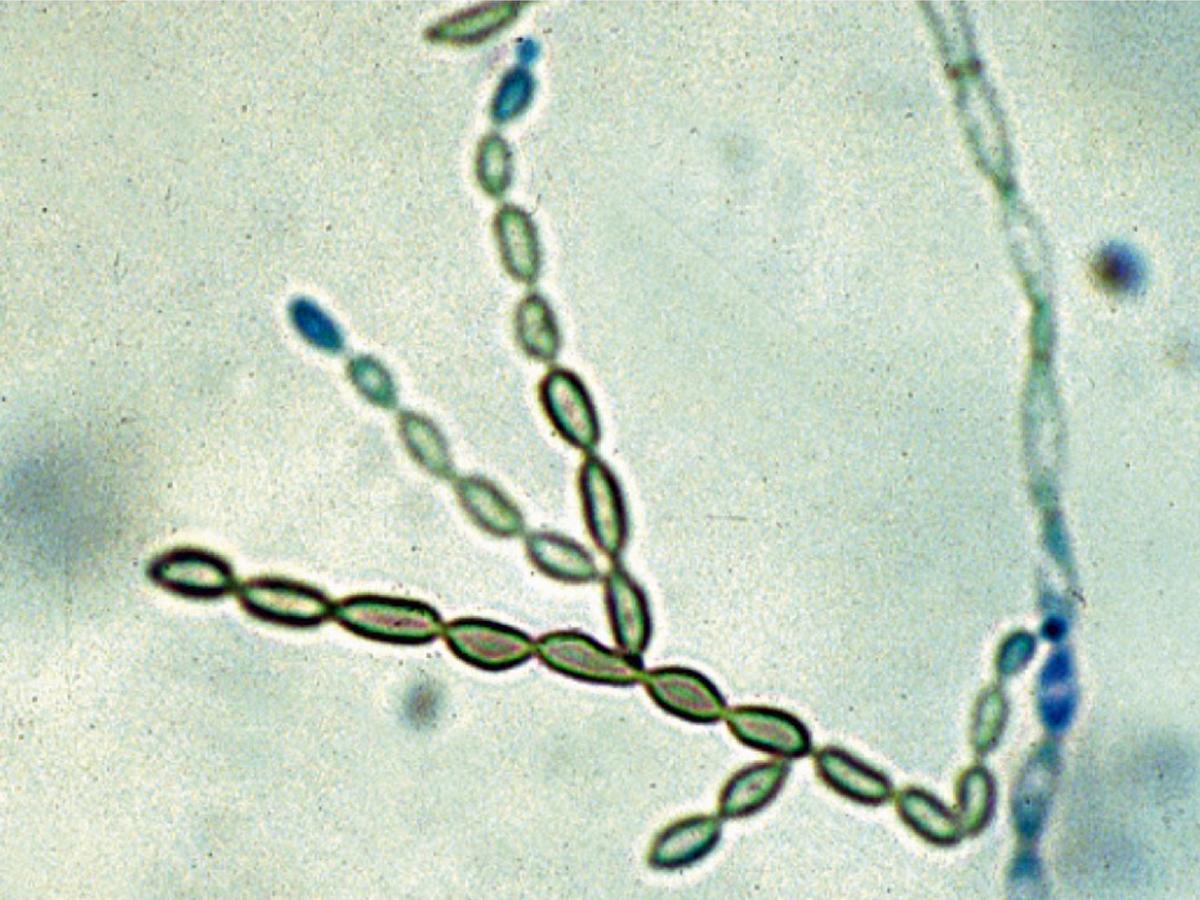
Cladophialophora bantiana conidiophore and conidia.
C. bantiana may be distinguished from Cladosporium species by the absence of conidia with distinctly pigmented hila, the absence of shield cells and by growth at >40C (compared with C. carrionii which has a maximum growth temperature of 35-37C, and Cladosporium species which have a maximum of <35C). C. bantiana may be further distinguished from C. carrionii by the formation of very long, sparsely branched chains of conidia.
Molecular identification:
ITS sequencing is recommended (Gerrits van den Ende and de Hoog, 1999; Badali et al. 2010).References:
McGinnis (1980), McGinnis and Borelli (1981), McGinnis et al. (1986a), Rippon (1988), Kwon-Chung and Bennett (1992), de Hoog et al. (2000, 2015), Chakrabarti et al. (2016).Antifungal susceptibility: Cladophialophora bantiana limited data available (Australian national data); MIC µg/mL. No ≤0.016 0.03 0.06 0.125 0.25 0.5 1 2 4 ≥8 AmB 27 1 3 2 6 6 9 ISAV 3 2 1 VORI 27 3 3 7 11 3 POSA 25 7 5 12 1 ITRA 27 6 3 5 11 2 -
Cladophialophora carrionii
Synonym:
Cladosporium carrioniiCladophialophora carrionii is a recognised agent of chromoblastomycosis and it has been isolated from soil and fence posts made from Eucalyptus spp. Cases of chromoblastomycosis caused by C. carrionii are commonly found in Australia, Venezuela, Madagascar and South America. Isolates from phaeomycotic cysts and opportunistic infections have also been reported.
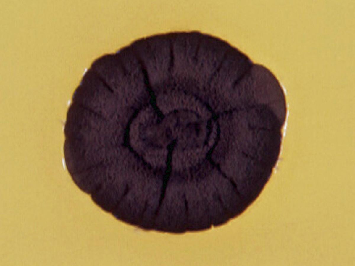
Culture of Cladophialophora carrionii.
RG-2 organism.
Morphological description:
Colonies are slow growing, reaching 3-4 cm in diameter after one month, with a compact suede-like to downy surface and are olivaceous-black in colour. Microscopy shows ascending to erect, olivaceous-green, apically branched, elongate conidiophores producing branched acropetal chains of conidia. Conidia are pale olivaceous, smooth-walled or slightly verrucose, limoniform to fusiform, 1.5-3.0 x 2.0-7.0 µm in size. Bulbous phialides with large collarettes and minute, hyaline conidia are occasionally formed on nutritionally poor media. Maximum growth temperature 35-37C.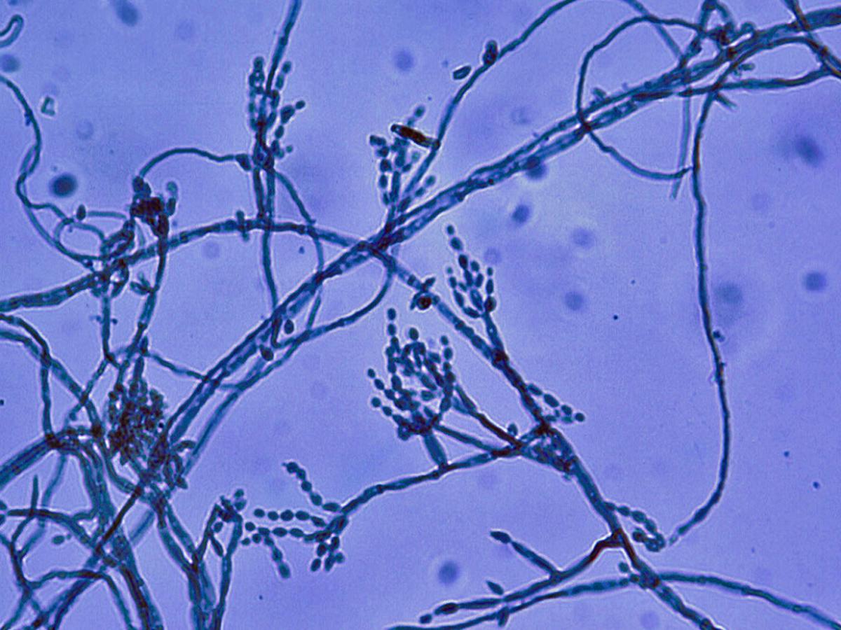
Conidiophores and conidia of C. carrionii.
Molecular identification:
ITS sequencing is recommended (Abliz et al. 2004; de Hoog et al. 2007).Key features:
Conidia are smaller and comprise heavily branched chains which fall apart much more easily than in the other Cladophialophora species.References:
McGinnis (1980), Rippon (1988), de Hoog et al. (1995, 2000, 2015).Antifungal susceptibility: Cladophialophora carrionii limited data available (Australian national data); MIC µg/mL. No ≤0.008 0.016 0.03 0.06 0.125 0.25 0.5 1 2 4 ≥8 AmB 2 1 1 VORI 2 1 1 POSA 2 1 1 ITRA 2 1 1 Antifungal susceptibility: Cladophialophora carrionii limited data available (McGinnis and Pasarell 1998a, 1998b, Espinel-Ingroff et al. 2001, Gonzales et al. 2005); MIC µg/mL. Antifungal Range MIC90 Antifungal Range MIC90 AmB 0.06-4 1 VORI 0.03-0.5 0.25 ITRA 0.06-0.5 0.125 POSA 0.06-0.5 0.25 TERB 0.03-0.125 Geometric mean = 0.03