Rhinocladiella
Rhinocladiella contains six to eight species, with five species of medical interest; R. aquaspersa, R. atrovirens, R. basitona, R. mackenziei (formerly Ramichloridium mackenziei) and R. similis. R. mackenziei is a frequently fatal neurotropic organism and appears to be restricted to individuals residing in, or immigrating from, Middle Eastern countries (Revankar and Sutton 2010).
Species descriptions
-
Rhinocladiella atrovirens complex
Morphologically Rhinocladiella atrovirens is identical to R. similis, but the species are phylogenetically distinct. In the past, some cases reported under the name R. atrovirens may have concerned R. similis (Revankar and Sutton, 2010; Taj-Aldeen et al., 2010).
RG-1 organism.
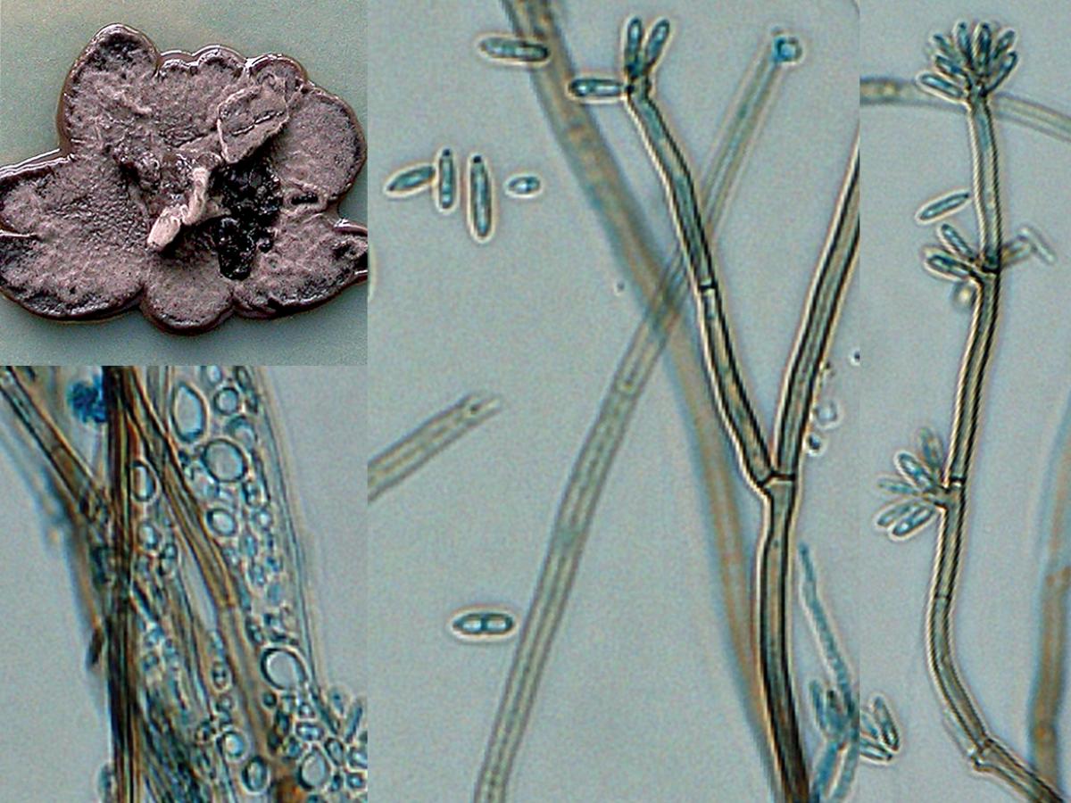
Culture, conidiophores showing a terminal denticulate rachis, conidia and budding yeast cells of Rhinocladiella atrovirens.
Morphological description:
Colonies are restricted, velvety or lanose, olivaceous, often slightly mucoid at the centre; reverse dark olivaceous green to blackish. Conidiophores are short, brown, thick-walled. Conidiogenous cells are cylindrical, intercalary or free, 9-19 x 1.6-2.2 µm; denticulate rachis up to 15 µm long, with crowded, flat or butt-shaped, unpigmented conidial denticles. Conidia are hyaline, thin- and smooth-walled, short-cylindrical, with truncate basal scars, 3.7-5.5 x 1.2-1.8 µm. Budding cells, if present, are hyaline, thin-walled, broadly ellipsoidal, 3.0-4.3 x 1.7-2.5 µm. Germinating cells are inflated, spherical to subspherical, 4.5-6.0 µm. An annellidic Exophiala synanamorph may be present.Molecular identification:
ITS sequencing is recommended for accurate species identification (Taj-Aldeen et al. 2010).References:
de Hoog (1977, 1983), Schell et al. (1983), de Hoog et al. (2000, 2015).Antifungal susceptibility: Rhinocladiella atrovirens very limited data (Australian national data); MIC µg/mL.
No ≤0.03 0.06 0.125 0.25 0.5 1 2 4 8 ≥16 AmB 2 1 1 ISAV 1 1 VORI 2 1 1 POSA 2 1 1 ITRA 2 2 -
Rhinocladiella mackenziei complex
WARNING: RG-3 organism.
Cultures of R. mackenziei represent a potential biohazard to laboratory personnel and must be handled with extreme caution in a Class II Biological Safety Cabinet (BSCII).R. mackenziei is an extremely rare, neurotropic organism that causes fatal brain lesions, mostly in patients who are immunocompromised or suffered from underlying metabolic diseases. The species is typically restricted to the Middle East, in an arid zone between Israel and Pakistan (Kanj et al. 2001, Khan et al. 2002, Taj-Aldeen et al. 2010), with a single autochtonous case in India (Badali et al. 2010). Cases in the USA and Europe invariably were found in patients originating from the Middle East (Sutton et al. 1998, Revankar and Sutton 2010, de Hoog et al. 2015).
Morphological description:
Colonies growing moderately rapidly, velvety, olivaceous-brown. Conidiophores arising at right angles from creeping hyphae, stout, thick-walled, brown, 3.0-4.5 µm wide, 10-25 µm long, apically with short-cylindrical denticles. Conidia brown, ellipsoidal, 8.5-12.0 × 4-5 µm, with a prominent, wide basal scar.Molecular identification:
ITS and D1/D2 sequencing is recommended for accurate species identification (Taj-Aldeen et al. 2010).References:
de Hoog (1977, 1983), Schell et al. (1983), de Hoog et al. (2000, 2015), Taj-Aldeen et al. (2010), Revankar and Sutton (2010).Antifungal susceptibility: Rhinocladiella mackenziei limited data (Australian national data); MIC µg/mL.
No ≤0.016 0.03 0.06 0.125 0.25 0.5 1 2 4 ≥8 AmB 7 1 3 3 VORI 3 1 1 1 POSA 5 2 2 1 ITRA 7 5 1 1