Stemphylium
Most species of Stemphylium are plant pathogens with occasional isolates from soil, they are rarely seen in the clinical laboratory.
RG-1 organism.
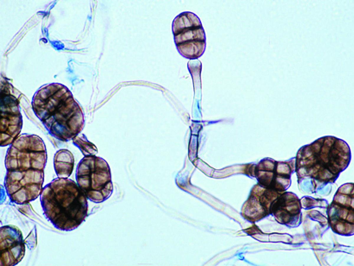
Conidiophore and conidia of Stemphylium spp.
Morphological description:
Colonies are rapid growing, brown to olivaceous-black or greyish and suede-like to floccose. Microscopically, solitary, darkly pigmented, terminal, multicellular conidia (dictyoconidia) are formed on a distinctive conidiophore with a darker terminal swelling. Note: The conidiophore proliferates percurrently through the scar where the terminal conidium (poroconidium) was formed. Conidia are pale to mid-brown, oblong, rounded at the ends, ellipsoidal, obclavate or subspherical and are smooth or in part verrucose. Stemphylium should not be confused with Ulocladium which produces similar dictyoconidia from a sympodial conidiophore, not from a percurrent conidiogenous cell as in Stemphylium.
Molecular identification:
ITS sequencing (Woudenberg et al. 2013).
Key features:
Dematiaceous hyphomycete producing darkly pigmented, dictyoconidia from the swollen end of a percurrent conidiophore.
References:
Ellis (1971, 1976), Rippon (1988), de Hoog et al. (2000).