Chrysosporium
Species of Chrysosporium are occasionally isolated from skin and nail scrapings, especially from feet, but because they are common soil saprophytes they are usually considered contaminants.
There are about 70 species of Chrysosporium, several are keratinolytic with some also being thermotolerant, and cultures may closely resemble some dermatophytes, especially Trichophyton mentagrophytes. Some strains may also resemble cultures of Histoplasma and Blastomyces.
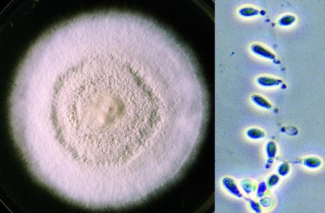
Culture of Chrysosporium tropicum showing typical pyriform to clavate shaped conidia with truncated bases which may be formed either intercalary, laterally or terminally.
Morphological description:
Colonies are moderately fast growing, flat, white to tan to beige in colour, often with a powdery or granular surface texture. Reverse pigment absent or pale brownish-yellow with age. Hyaline, one-celled conidia are produced directly on vegetative hyphae by non-specialised conidiogenous cells. Conidia are typically pyriform to clavate with truncate bases and are formed either intercalary (arthroconidia), laterally (often on pedicels) or terminally.
Molecular identification:
Chrysosporium is phylogenetically heterogeneous; the polyphyletic origin of the genus was first demonstrated by Vidal et al. (2000) on the basis of ITS sequences, and further elaborated by Stchigel et al. (2014). ITS sequencing can assist in identification of clinical isolates.
Chrysosporium tropicum
RG-2 organism.
Morphological descriptions:
Colonies are flat, white to cream-coloured with a very granular surface. Reverse pigment absent or pale brownish-yellow with age. Microscopically, conidia are numerous, hyaline, single-celled, clavate to pyriform, smooth, slightly thick-walled (6-7 x 3.5-4 µm), and have broad truncate bases and pronounced basal scars. The conidia are formed at the tips of the hyphae, on short or long lateral branches, or sessile along the hyphae (intercalary). No macroconidia or hyphal spirals are seen.
References:
Carmichael (1962), Rebell and Taplin (1970), Sigler and Carmichael (1976), van Oorschot (1980), Domsch et al. (2007), de Hoog et al. (2000, 2015).