Phialophora verrucosa
The genus Phialophora contains more than 40 species, most are saprophytes commonly found in soil or on decaying wood.
Some human pathogens with phialidic conidiogenesis previously assigned to Phialophora have been moved to other genera, namely, Phaeoacremonium and Pleurostomophora. P. verrucosa, P. americana, P. bubakii, P. europaea and P. reptans remain of medical interest (Revankar and Sutton 2010). Both P. verrucosa and P. americana produce their conidia from phialides with conspicuous darkened collarettes, however sequencing has demonstrated a close relatedness, suggesting that these species may be synonymous (de Hoog et al. 1999). P. verrucosa is primarily an agent of chromoblastomycosis although other reported infections include endocarditis, keratitis, and osteomyelitis.
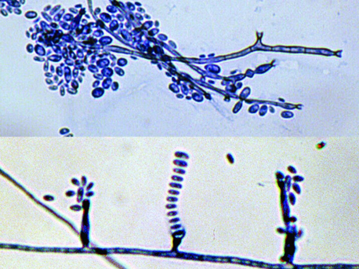
Phialides and conidia of P. verrucosa.
RG-2 organism.
Morphological description:
Colonies (SDA) are slow growing, initially dome-shaped, later becoming flat, suede-like and olivaceous to black in colour. Phialides are flask-shaped or elliptical with distinctive funnel-shaped, darkly pigmented collarettes. Conidia are ellipsoidal, smooth-walled, hyaline, mostly 3.0-5.0 x 1.5-3.0 μm, and aggregate in slimy heads at the apices of the phialide.
Key features:
Characteristic flask-shaped phialides with distinctive funnel-shaped, darkly pigmented collarettes.
Molecular identification:
ITS sequencing recommended (de Hoog et al. 1999).
References:
Ellis (1971), McGinnis (1978a, 1980), Domsch et al. (1980), de Hoog et al. (1999, 2000, 2015), Revankar and Sutton (2010).
|
Antifungal susceptibility: Phialophora verrucosa limited data (Australian national data); MIC µg/mL. |
||||||||||||
|---|---|---|---|---|---|---|---|---|---|---|---|---|
| No | ≤0.03 | 0.06 | 0.125 | 0.25 | 0.5 | 1 | 2 | 4 | 8 | 16 | ≥32 | |
| AmB | 7 | 1 | 4 | 2 | ||||||||
| VORI | 7 | 1 | 3 | 3 | ||||||||
| POSA | 5 | 2 | 4 | 1 | ||||||||
| ITRA | 7 | 1 | 4 | 2 | ||||||||