Cryptococcus
The genus Cryptococcus is characterised by globose to elongate yeast-like cells or blastoconidia that reproduce by narrow-necked budding.
Pseudohyphae are absent or rudimentary. Most species are encapsulated, although the extent of capsule formation depends on the medium.
Under certain conditions of growth, the capsule may contain starch-like compounds, which are released into the medium by many strains. Within tissue sections, mucicarmine or Alcian blue stains the capsule of Cryptococcus species to distinguish it from other yeasts with similar morphologies.
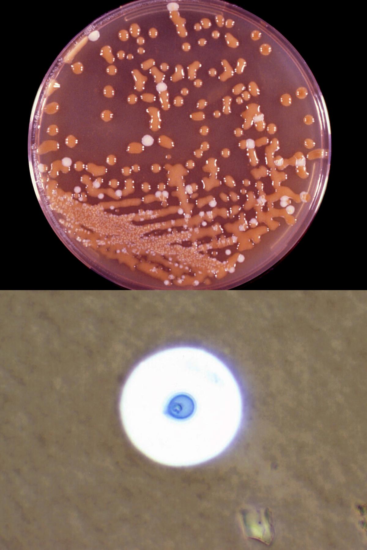
Bird seed agar plate showing brown colonies of C. neoformans and white colonies of Candida albicans and India ink preparation showing capsules of C. neoformans.
On solid media the cultures are generally mucoid or slimy in appearance; red, orange or yellow carotenoid pigments may be produced, but young colonies of most species are usually non-pigmented, and cream in colour. All Cryptococcus species produce urease and are non-fermentative. Nitrate may be assimilated or not; inositol assimilated. The genus Cryptococcus differs from the genus Rhodotorula in its inositol assimilation.
Cryptococcosis is a chronic, subacute to acute pulmonary, systemic or meningitic disease, initiated by the inhalation of infectious propagules (basidiospores and/or desiccated yeast cells) from the environment. Primary pulmonary infections have no diagnostic symptoms and are usually subclinical. On dissemination, the fungus usually shows a predilection for the central nervous system, however skin, bones and other visceral organs may also become involved.
Cryptococcus neoformans and C. gattii are the principle pathogenic species. Naganishia albida (formerly Cryptococcus albidus) and Patiliotrema laurentii (formely Cryptococcus laurentii) have on occasion also been implicated in human infection.
Molecular identification:
Requires ITS and/or D1/D2 sequencing, particularly for identification of unusual species.
MALDI-TOF MS:
Can provide reliable species and subspecies level identification of Cryptococcus species, but its accuracy is dependent on database quality (Arendrup et al. 2014).
References:
Rippon (1982), Barnett et al. (1983), Kurtzman et al. (2011), Casadevall and Perfect (1998), de Hoog et al. (2000, 2015), McTaggart et al. (2013).
Species descriptions
-
Cryptococcus gattii
Synonymy:
Filobasidiella bacillispora; Cryptococcus neoformans var. gattiiRG-2 organism.
Cryptococcus gattii has two serotypes (B and C) and was reclassified as a separate species from C. neoformans in 2002 (Kwon-Chung et al. 2002). C. gattii generally has a more restricted geographical distribution than C. neoformans, causing human disease in climates ranging from temperate to tropical Australia, Papua New Guinea, parts of Africa, India, Southeast Asia, Mexico, Brazil, Paraguay and Southern California, although recent infections have also been reported from Vancouver Island, Canada and in the Pacific Northwest, USA (Pfaller & Diekema, 2010, Espinel-Ingroff and Kidd, 2015). C. gattii has a specific ecological association with numerous species of Eucalyptus trees, although the Canadian isolates are associated with a range of native non-Eucalyptus species (Kidd et al. 2007). Historically considered a pathogen in immunocompetent hosts, a recent review in Australia noted an increase in C. gattii infections in HIV-negative immunocompromised patients (Chen et al.2012). Cryptococcosis caused by C. gattii is often associated with large mass lesions (cryptococcomas) in the lung and/or brain (Sorrell, 2001).
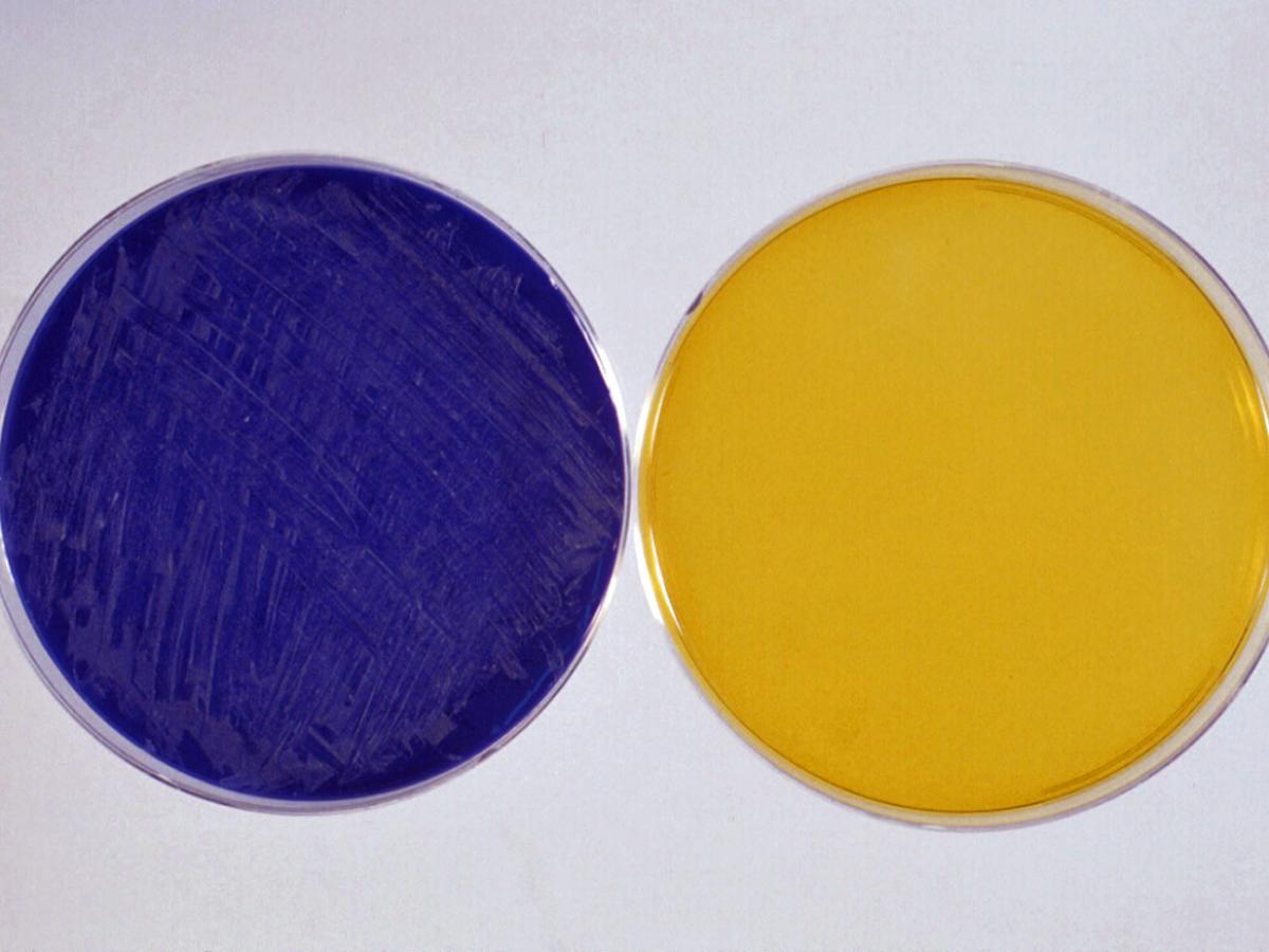
Cryptococcus gattii turns CGB agar blue within 2-5 days; Cryptococcus neoformans does not grow on this medium.
Canavanine glycine bromothymol blue (CGB) agar:
Kwon-Chung et al. (1982) is the media of choice to differentiate C. gattii from C. neoformans. This simple biotype test is based on the ability of C. gattii isolates to grow in the presence of L-canavanine and to assimilate glycine as a sole carbon source. A heavy inoculum is important.Culture:
Colonies (SDA) cream-coloured smooth, mucoid, yeast-like colonies.Microscopy:
Globose to ovoid budding yeast-like cells 3.0-7.0 x 3.3- 7.9 µm.India ink preparation:
Positive - distinct, wide gelatinous capsules are present. Some strains may not produce apparent capsules from culture.Dalmau plate culture:
Budding yeast cells only. No pseudohyphae present.Bird seed agar:
Colonies turn dark brown in colour as colonies selectively absorb a brown pigment from this media. Colonies are often more mucoid when compared with C. neoformans (Staib, 1987).Canavanine glycine bromothymol blue (CGB) agar:
Turns blue within 2-5 days.Physiological Tests: + Positive, - Negative, v Variable, w Weak, s Slow, nd No Data Germ Tube - L-Sorbose - L-Arabinose +,w D-Glucitol + Fermentation Sucrose + D-Arabinose + 𝝰-M-D-Glucoside + Glucose - Maltose + D-Ribose v D-Gluconate + Galactose - Cellobiose +,w L-Rhamnose + DL-Lactate - Sucrose - Trehalose + D-Glucosamime v myo-Inositol + Maltose - Lactose - N-A-D-glucosamine v 2-K-D-Gluconate nd Lactose - Melibiose - Glycerol - D-Glucuronate nd Trehalose - Raffinose +,w Erythritol - Nitrate - Assimilation Melezitose + Ribitol v Urease + Glucose + Soluble Starch + Galactitol + 0.1% Cycloheximide - Galactose + D-Xylose + D-Mannitol + Growth at 37C + Key features:
Encapsulated yeast; absence of pseudohyphae; growth at 37C; positive hydrolysis of urea; negative fermentation of sugars and positive assimilation of glucose, maltose, sucrose, galactose, trehalose, raffinose, inositol, cellobiose, rhamnose, arabinose, melezitose and xylose, and negative assimilation of nitrate, lactose, melibiose, erythritol and soluble starch; growth on bird seed (Guizotia abyssinica seed) or caffeic acid agar - colonies turn a dark brown colour; growth on CGB agar turning it blue within 2-5 days.Antifungal susceptibility: Cryptococcus gattii (Espinel-Ingroff et al. 2015 and Australian national data); MIC µg/mL. Note: All Cryptococcus species are intrinsically resistant to echinocandins.
Antifungal No ≤0.016 0.03 0.06 0.125 0.25 0.5 1 2 4 8 16 32 ≥64 AMB 271 1 3 18 52 61 50 81 5 FLU 271 1 5 27 55 82 63 32 5 1 ISAV 456 118 111 98 96 26 7 VORI 249 36 55 79 46 28 5 POSA 209 19 21 44 62 49 12 1 1 ITRA 271 23 32 64 97 51 4 5FC 271 3 18 75 102 58 11 4 -
Cryptococcus neoformans
Synonymy:
Filobasidiella neoformans; Cryptococcus neoformans var. neoformans.RG-2 organism.
This species comprises two varieties: C. neoformans var. grubii (serotype A) and C. neoformans var. neoformans (serotype D).
C. neoformans var. grubii:
Has a worldwide distribution, causing 95% of all C. neoformans infections. It has been isolated from various sources in nature and is noted for its association with accumulations of avian guano, especially with pigeon excreta. The fungus has also been isolated from the dung of caged birds including canaries, parrots and budgerigars. Other environmental isolations of C. neoformans var. grubii include rotting vegetables, fruits and fruit juices, wood, dairy products and soil.C. neoformans var. neoformans:
Has a more restricted distribution with infections being more prevalent in Europe, including France, Italy and Denmark, where it accounts for 30% of isolates. Moreover, C. neoformans var. neoformans infections are more strongly correlated with older patients, the skin, and the use of corticosteroids (Franzot et al. 1999).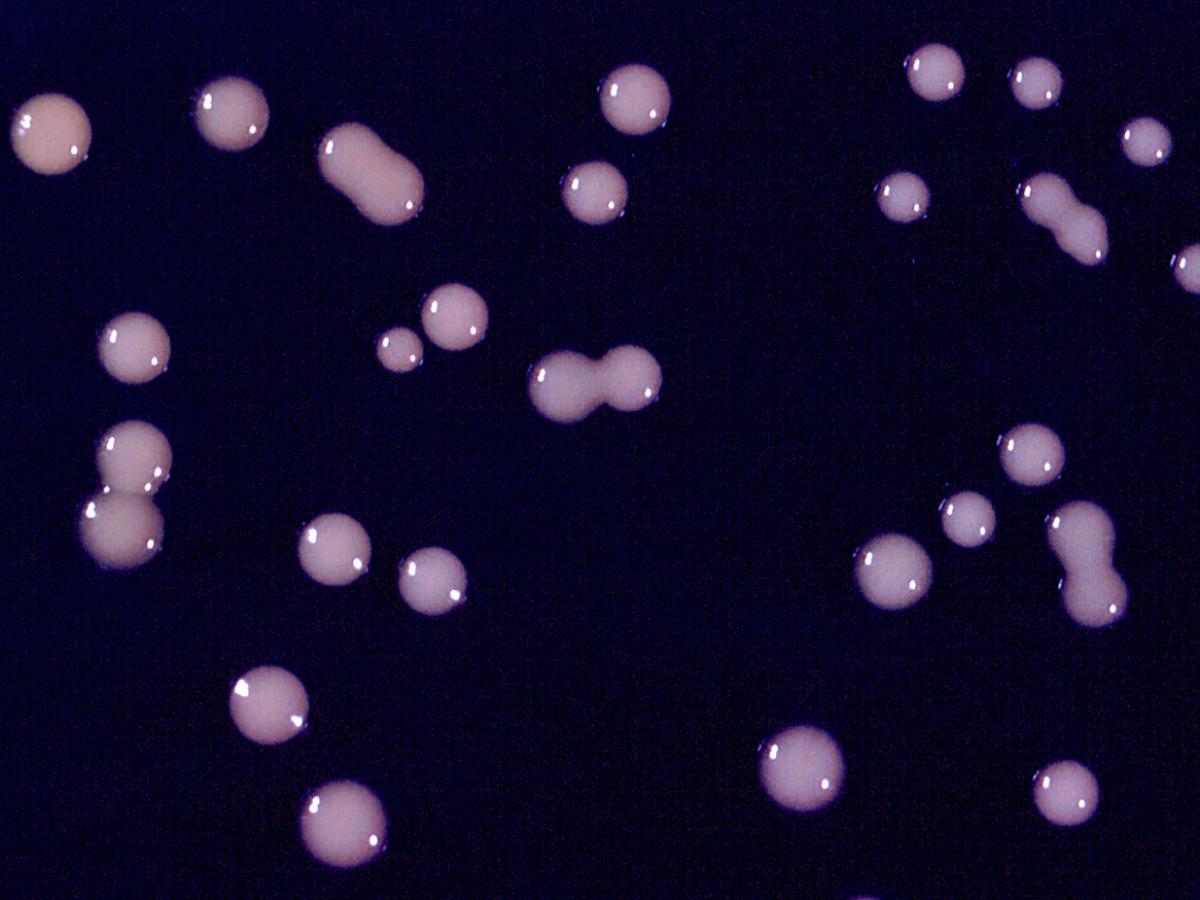
Culture of C. neoformans. Note: mucoid colonies.
Culture:
Colonies (SDA) cream-coloured smooth, mucoid, yeast-like colonies.Microscopy:
Globose to ovoid budding yeast-like cells 3.0-7.0 x 3.3-7.9 µm.India ink preparation:
Positive - distinct, wide gelatinous capsules are present on direct microscopy. Some strains may not produce apparent capsules from culture.Dalmau plate culture:
Budding yeast cells only. No pseudohyphae present.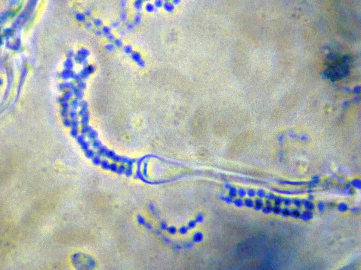
Basidium with basidiospores of C. neoformans.
Bird seed agar: Colonies turn dark brown in colour as colonies selectively absorb a brown pigment from this media (Staib, 1987).
Canavanine glycine bromothymol blue (CGB) agar:
No growth or colour change.Creatinine dextrose bromothymol blue thymine (CDBT) agar:
May be used to differentiate C. neoformans var. neoformans and C. neoformans var. grubii. C. neoformans var. neoformans grows as bright red colonies, turning the medium a bright orange after 5 days while no colour change is observed for C. neoformans var. grubii (Irokanulo et al. 1994).Physiological Tests: + Positive, - Negative, v Variable, w Weak, s Slow, nd No Data Germ Tube - L-Sorbose - L-Arabinose +,w D-Glucitol + Fermentation Sucrose + D-Arabinose + 𝝰-M-D-Glucoside + Glucose - Maltose + D-Ribose v D-Gluconate + Galactose - Cellobiose +,w L-Rhamnose + DL-Lactate - Sucrose - Trehalose + D-Glucosamime v myo-Inositol + Maltose - Lactose - N-A-D-glucosamine v 2-K-D-Gluconate nd Lactose - Melibiose - Glycerol - D-Glucuronate nd Trehalose - Raffinose +,w Erythritol - Nitrate - Assimilation Melezitose + Ribitol v Urease + Glucose + Soluble Starch + Galactitol + 0.1% Cycloheximide - Galactose + D-Xylose + D-Mannitol + Growth at 37C + Key features:
Encapsulated yeast; absence of pseudohyphae; growth at 37C; positive hydrolysis of urea; negative fermentation of sugars and positive assimilation of glucose, maltose, sucrose, galactose, trehalose, raffinose, inositol, cellobiose, rhamnose, arabinose, melezitose and xylose, and negative assimilation of nitrate, lactose, melibiose, erythritol and soluble starch; growth on bird seed (Guizotia abyssinica seed) or caffeic acid agar - colonies turn a dark brown colour; does not grow on CGB agar (no colour change).Antifungal susceptibility: Cryptococcus neoformans (Espinel-Ingroff et al. 2015, Australian National data); MIC µg/mL. Note: All Cryptococcus species are intrinsically resistant to echinocandins.
Antifungal No ≤0.016 0.03 0.06 0.125 0.25 0.5 1 2 4 8 16 32 ≥64 AMB 522 2 5 17 85 64 137 174 37 1 FLU 522 1 2 30 67 108 149 118 35 11 1 ISAV 584 235 206 110 23 8 2 VORI 484 132 144 123 62 22 1 POSA 439 24 67 114 152 70 12 ITRA 523 36 99 159 174 48 6 1 5FC 523 2 1 15 50 86 130 165 63 8 1 2