Trichosporon
The taxonomy of Trichosporon has been redefined: Trichosporon cutaneum, T. dermatis, T. jirovecii and T. mucoides have now been transferred to the new genus Cutaneotrichosporon, while Trichosporon domesticum, T. loubieri and T. mycotoxinovorans have now been included into the re-defined genus Apiotrichum (Liu et al. 2015).
Trichosporon species are urease-positive, non-encapsulated basidiomycetous yeasts characterised by the development of hyaline, septate hyphae that fragment into oval or rectangular arthroconidia. Some blastoconidia are also seen. The colonies are usually raised and have a waxy appearance, which develop radial furrows and irregular folds. They are widely distributed in the environment many have different habitats and usually occupy narrow ecologic niches. Some are soil borne and others are associated with humans and animals (Guarro et al. 1999, Sugita 2011, Arendrup et al. 2015).
Four species Trichosporon asahii, T. asteroides, T. inkin, and T. ovoides are the most common clinical isolates, however, T. coremiiforme, T. dohaense ,T. faecale, T. japonicum and T. lactis have also been reported from human and animal infections (Guarro et al. 1999; Rodriguez-Tudela et al. 2005, Chagas-Neto et al. 2008). Importantly, all species are resistant to echinocandins (Arendrup et al. 2014).
Trichosporon species are a minor component of normal skin flora, and are widely distributed in nature. They are regularly associated with the soft nodules of white piedra, and have been involved in a variety of opportunistic infections in the immunosuppressed patient. Disseminated infections are most frequently (75%) caused by T. asahii (Arendrup et al. 2014) and have been associated with leukaemia, organ transplantation, multiple myeloma, aplastic anaemia, lymphoma, solid tumours and AIDS. Disseminated infections are often fulminate and widespread, with lesions occurring in the liver, spleen, lungs and gastrointestinal tract. Infections in non-immunosuppressed patients include endophthalmitis after surgical extraction of cataracts, endocarditis usually following insertion of prosthetic cardiac valves, peritonitis in patients on continuous ambulatory peritoneal dialysis (CAPD), and intravenous drug abuse.
Note:
Genus identification is mandatory for clinical management and should be performed and provided in a timely manner. Species identification remains difficult and requires molecular analysis or MALDI-TOF MS (with an extensive database) (Arendrup et al. 2014).
Molecular identification:
ITS and D1/D2 sequencing is required for accurate species identification (Arendrup et al. 2014).
MALDI-TOF MS:
A promising identification tool to accurately identify species (with an extensive database) (Kolecka et al. 2013).
Comment:
The API 20C yeast identification system may be used for sugar assimilation tests.
References:
Kurtzman and Fell (1988), Gueho et al. (1992), de Hoog et al. (2000, 2015), Rodriguez-Tudela et al. (2005), Chagas-Neto et al. (2008), Guo et al. (2011), Xiao et al. (2013).
| Basic phenotypic key to medically important species. | ||
|---|---|---|
| 1 | Growth with melibiose No growth with melibiose |
2 3 |
| 2 | Tolerant to cycloheximide Not tolerant to cycloheximide |
Cutaneotrichosporon mucoides Cutaneotrichosporon cutaneum |
| 3 |
Growth with myo-inositol, no growth with L-arabinose |
Trichosporon inkin 4 |
| 4 | Colony very slow growth; thallus consisting of clumps of meristematic cells Colonies and microscopy otherwise |
Trichosporon asteroides 5 |
| 5 | Appressoria present in slide cultures Appressoria absent in slide cultures |
Trichosporon ovoides 6 |
| 6 | Arthroconidia barrel-shaped; thallus not meristematic Arthroconidia elongate, or thallus meristematic |
Trichosporon asahii Trichosporon asteroides |
Species descriptions
-
Trichosporon asahii
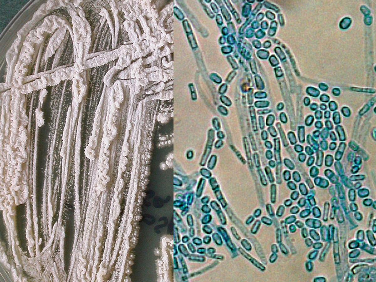
Trichosporon asahii culture and hyphae and arthroconidia.
RG-2 organism.
Morphological description:
Colonies are white to cream-coloured, powdery, suede-like to farinose with radial furrows and irregular folds. Budding cells and lateral conidia are absent. Arthroconidia are barrel-shaped. Appressoria absent. This species assimilates L-arabinose but not melibiose. Growth at 37C. Most common species, especially from invasive infections.Physiological Tests: + Positive, - Negative, v Variable, w Weak, s Slow, nd No Data Glucose + Melibiose - L-Rhamnose + D-Glucitol v Galactose + Raffinose - D-Glucosamime + 𝝰-M-D-Glucoside + L-Sorbose v Melezitose v N-A-D-glucosamine + D-Gluconate + Sucrose v Soluble Starch v Glycerol v DL-Lactate v Maltose + D-Xylose v Erythritol + myo-Inositol v Cellobiose + L-Arabinose + Ribitol v Nitrate - Trehalose + D-Arabinose + Galactitol - 2-K-D-Gluconate + Lactose + D-Ribose + D-Mannitol v D-Glucuronate + Antifungal susceptibility: Trichosporon asahii (Australian national data); MIC µg/mL.
Antifungal No ≤0.03 0.06 0.125 0.25 0.5 1 2 4 8 16 32 ≥64 AMB 56 4 4 16 18 15 2 FLU 56 1 7 26 20 2 ISAV 18 1 3 9 4 1 VORI 54 4 15 25 9 1 POSA 53 2 4 32 12 3 ITRA 56 1 8 40 6 1 -
Trichosporon asteroides
RG-2 organism.
Morphological description:
Colonies are restricted, dry, cream-coloured, cerebriform, with radial furrows and irregular folds. The meristematic form is punctiform, brownish and consists of hyphae which swell and become multiseptate which may fall apart into smaller packets. Budding cells and lateral conidia are absent. Arthroconidia are elongate and hyphae are often present. Appressoria absent. This species assimilates L-arabinose but not myo-inositol. Growth at 37C is variable. Uncommon species usually associated with superficial infections.Physiological Tests: + Positive, - Negative, v Variable, w Weak, s Slow, nd No Data Glucose + Melibiose - L-Rhamnose + D-Glucitol v Galactose + Raffinose - D-Glucosamime v 𝝰-M-D-Glucoside + L-Sorbose v Melezitose + N-A-D-glucosamine + D-Gluconate + Sucrose + Soluble Starch + Glycerol + DL-Lactate + Maltose + D-Xylose + Erythritol + myo-Inositol - Cellobiose + L-Arabinose + Ribitol v Nitrate - Trehalose + D-Arabinose + Galactitol - 2-K-D-Gluconate + Lactose + D-Ribose + D-Mannitol v D-Glucuronate + -
Trichosporon inkin
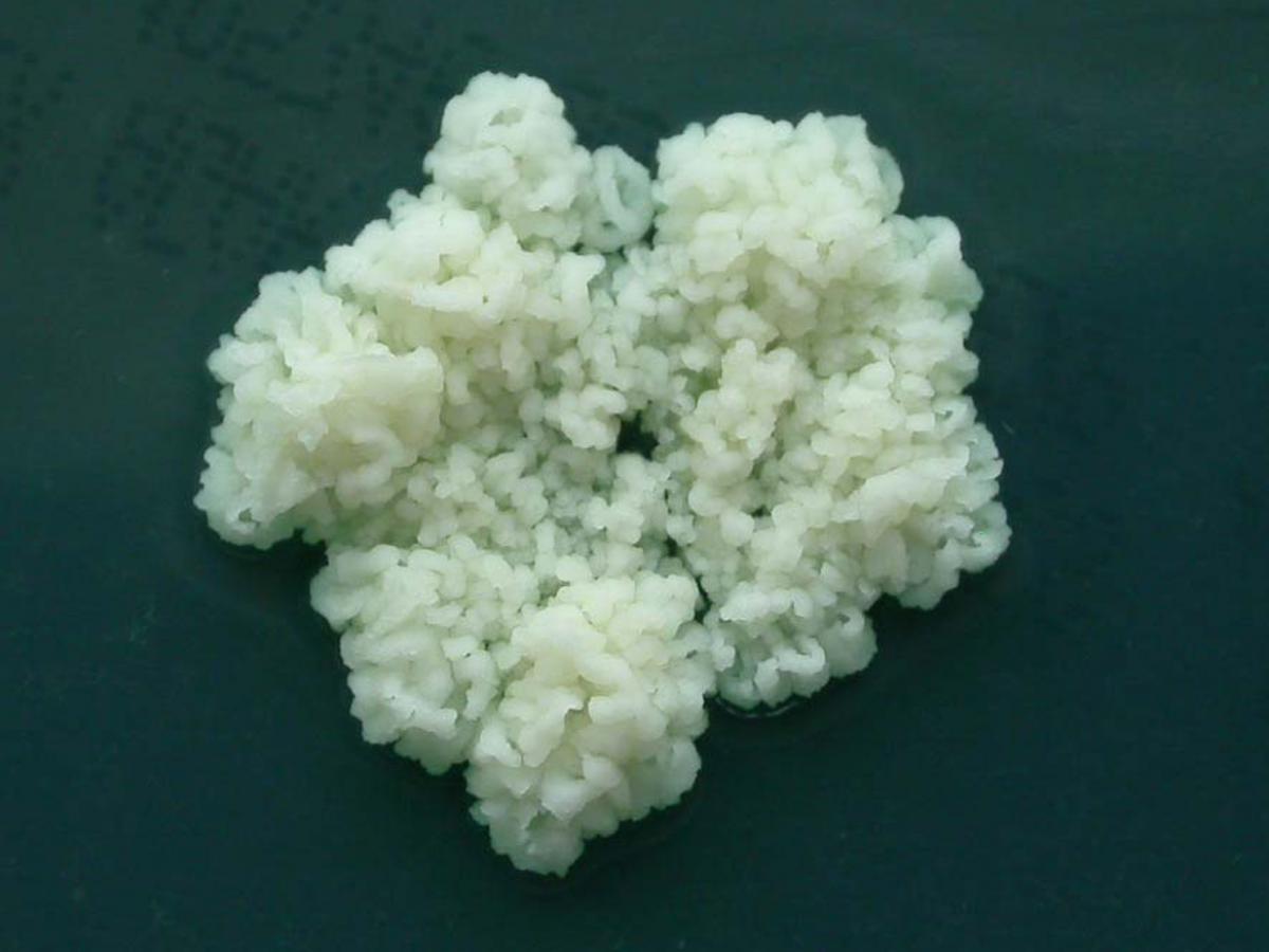
Trichosporon inkin colonies are restricted, white, finely cerebriform with a granular covering, without marginal zone, often cracking the media.
RG-2 organism.
Morphological description:
Colonies are restricted, white, finely cerebriform with a granular covering, without marginal zone, often cracking the media. Budding cells and lateral conidia absent. Arthroconidia are long cylindrical. Appressoria present in slide cultures. Sarcinae present on media with high sugar-content. This species assimilates myo-inositol but not melibiose and is tolerant to 0.01% (variable tolerance to 0.1%) cycloheximide. Growth at 37C. Usually associated with white piedra on pubic hairs.Physiological Tests: + Positive, - Negative, v Variable, w Weak, s Slow, nd No Data Glucose + Melibiose + L-Rhamnose - D-Glucitol + Galactose v Raffinose + D-Glucosamime v 𝝰-M-D-Glucoside + L-Sorbose v Melezitose + N-A-D-glucosamine + D-Gluconate + Sucrose + Soluble Starch + Glycerol v DL-Lactate + Maltose + D-Xylose + Erythritol + myo-Inositol + Cellobiose + L-Arabinose + Ribitol - Nitrate - Trehalose + D-Arabinose + Galactitol - 2-K-D-Gluconate + Lactose + D-Ribose + D-Mannitol v D-Glucuronate + Antifungal susceptibility: Trichosporon inkin (Australian national data); MIC µg/mL.
Antifungal No ≤0.03 0.06 0.125 0.25 0.5 1 2 4 8 16 32 ≥64 AMB 6 2 2 1 1 FLU 6 1 3 1 1 VORI 6 1 1 2 2 POSA 6 2 1 2 1 ITRA 6 2 2 2 -
Trichosporon ovoides
RG-2 organism.
Morphological description:
Colonies are restricted, white, granular, folded at the centre, with a flat marginal zone. Budding cells and lateral conidia absent. Arthroconidia are cylindrical. Appressoria present in slide cultures. This species does not assimilate melibiose, but tolerates 0.01% cycloheximide. Growth at 37C is variable. Uncommon species usually associated with superficial infections, like white piedra.Physiological Tests: + Positive, - Negative, v Variable, w Weak, s Slow, nd No Data Glucose + Melibiose - L-Rhamnose + D-Glucitol v Galactose + Raffinose v D-Glucosamime v 𝝰-M-D-Glucoside + L-Sorbose v Melezitose v N-A-D-glucosamine + D-Gluconate + Sucrose + Soluble Starch + Glycerol v DL-Lactate + Maltose + D-Xylose + Erythritol + myo-Inositol + Cellobiose + L-Arabinose v Ribitol - Nitrate - Trehalose v D-Arabinose v Galactitol - 2-K-D-Gluconate + Lactose + D-Ribose + D-Mannitol + D-Glucuronate +