Basidiobolus ranarum
Synonyms:
Basidiobolus meristosporus; Basidiobolus haptosporus; Basidiobolus heterosporus.
Basidiobolus ranarum is commonly present in decaying fruit and vegetable matter, and as a commensal in the intestinal tract of frogs, toads and lizards.
It has been reported from tropical regions of Africa and Asia including India, Indonesia and Australia.
RG-2 organism.
Morphological description:
Colonies are moderately fast growing at 30C, flat, yellowish-grey to creamy-grey, glabrous, becoming radially folded and covered by a fine, powdery, white surface mycelium. Satellite colonies are often formed by germinating conidia ejected from the primary colony. Microscopic examination usually shows the presence of large vegetative hyphae (8-20 µm in diameter) forming numerous round (20-50 µm in diameter), smooth, thick-walled zygospores that have two closely appressed beak-like appendages. The production of “beaked” zygospores is characteristic of the genus. Two types of asexual conidia are formed, although isolates often lose their ability to sporulate with subculture. Special media incorporating glucosamine hydrochloride and casein hydrolsate may be needed to stimulate sporulation (Shipton and Zahari, 1987). Primary conidia are globose, one-celled, solitary and are forcibly discharged from a sporophore. The sporophore has a distinct swollen area just below the conidium that actively participates in the discharge of the conidium. Secondary (replicative) conidia are clavate, one-celled and are passively released from a sporophore. These sporophores are not swollen at their bases. The apex of the passively released spore has a knob-like adhesive tip. These spores may function as sporangia, producing several sporangiospores.
Click images below to expand:
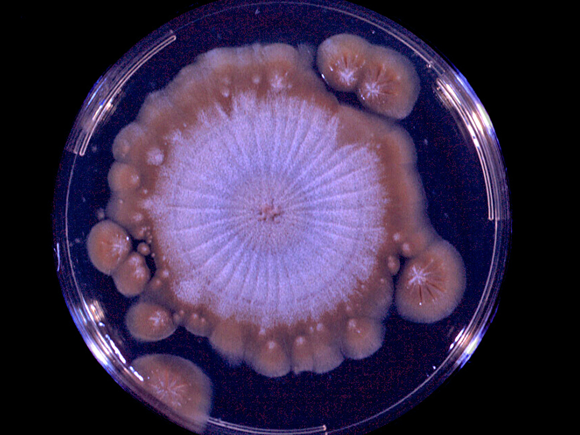
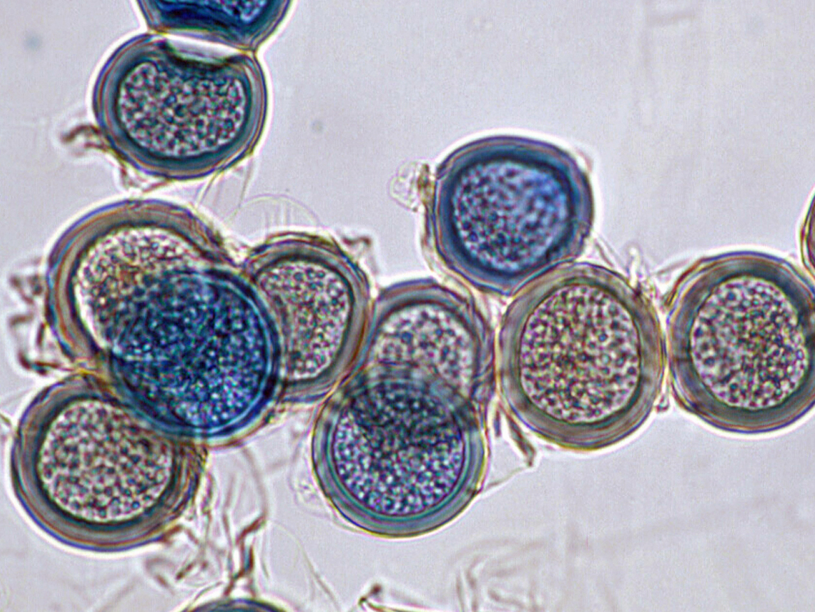
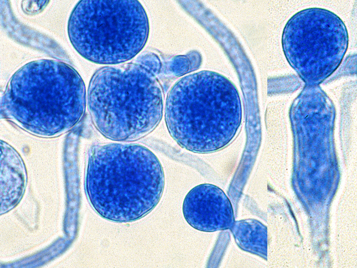
Images of Basidiololus ranarum showing a culture displaying satellite colonies that are often formed by germinating conidia ejected from the primary colony, "beaked" zygospores, conidia and a sporophore with a distinct swollen area just below the conidium.
References:
Strinivasan and Thirumalachar (1965), Greer and Friedman (1966), Dworzack et al. (1978), McGinnis (1980), King (1983), Rippon (1988), Davis et al. (1994), Jong and Dugan (2003), de Hoog et al. (2000, 2015) and Ellis (2005a).