Lichtheimia corymbifera
Synonymy:
Mycocladus corymbifera, Absidia corymbifera, Mucor corymbifera.
Recent taxonomic revisions of the genus Absidia have placed the thermotolerant species into the genus Lichtheimia (Hoffmann et al. 2007, 2009).
The genus Lichtheimia currently contains five mostly saprophytic plant decaying and soil-borne species. Lichtheimia corymbifera is the principle pathogen causing human and animal infections, however L. ramosa and L. ornata have also been reported as human pathogens (often misidentified morphologically as L. corymbifera) (Alastruey-Izquierdo et al. 2010).
RG-2 organism.
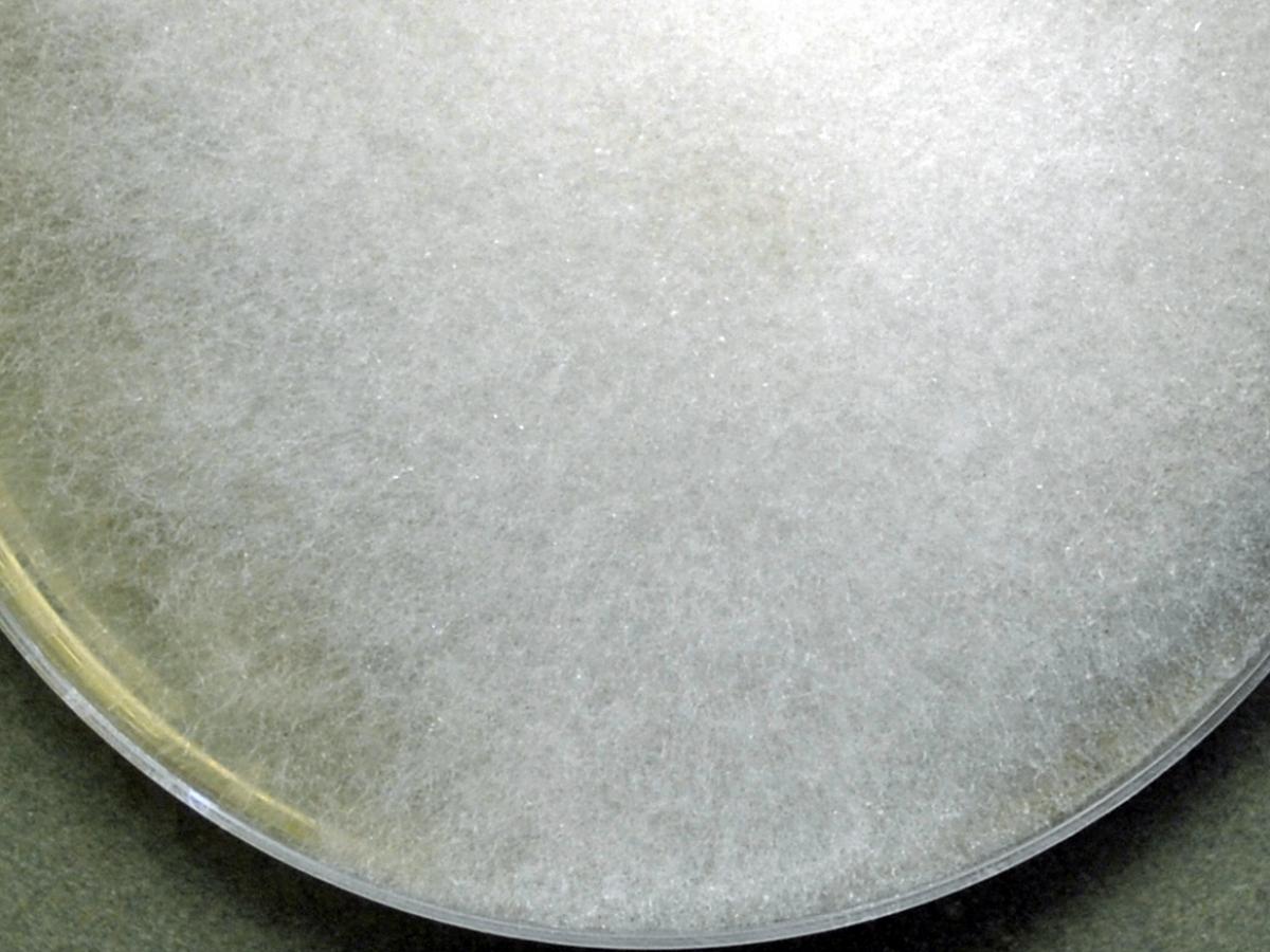
Culture of Lichtheimia corymbifera.
Morphological description:
Colonies are fast growing, floccose, white at first becoming pale grey with age, and up to 1.5 cm high. Sporangiophores are hyaline to faintly pigmented, simple or sometimes branched, arising solitarily or in groups. Sub-sporangial septa are absent or rare. Rhizoids are very sparingly produced and may be difficult to find without the aid of a dissecting microscope to examine the colony on the agar surface. Sporangia are small (10-40 µm in diameter) and are typically pyriform in shape with a characteristic conical-shaped columella and pronounced apophysis, often with a short projection at the top. Sporangiospores vary from subglobose to oblong-ellipsoidal (3-7 x 2.5-4.5 µm), hyaline to light grey and smooth-walled. Intercalary giant cells may also be present. Temperature: optimum 35-37C; maximum 46C.

Typical pyriform-shaped sporangium with a conical-shaped columella and pronounced apophysis of Lichtheimia corymbifera. Grocott methenamine silver (GMS) stained tissue section from a lung biopsy showing a typical sporangium of L. corymbifera.
Key features:
Mucorales, small pyriform-shaped sporangia with a characteristic conical-shaped columellae and pronounced apophyses, rapid growth at 40C.
Comment:
Morphological characteristics alone are not sufficient to reliably differentiate between L. corymbifera, L. ramosa and L. ornata. While L. ornata develops large, densely branched giant cells and L. ramosa has more ellipsoidal to cylindrical sporangiospores and a faster growth rate, these characters are often difficult to interpret. Molecular methods are needed to accurately separate these species.
Molecular identification:
Species recognition in Lichtheimia is based on ITS and/or D1/D2 sequencing (Garcia-Hermoso et al. 2009, Alastruey-Izquierdo et al. 2010).
MALDI-TOF MS:
Direct identification of Lichtheimia species was described by Schrödl et al. (2012).
References:
Ellis and Hesseltine (1965, 1966), Hesseltine and Ellis (1964a, 1966), Nottebrock et al. (1974), O’Donnell (1979), Samson et al. (1995), Domsch et al. (1980), McGinnis (1980), Ellis (2005b), de Hoog et al. (2000, 2015).
| Antifungal | No | ≤0.016 | 0.03 | 0.06 | 0.125 | 0.25 | 0.5 | 1 | 2 | 4 | 8 | 16 | ≥32 |
|---|---|---|---|---|---|---|---|---|---|---|---|---|---|
| AMB | 179 | 9 | 31 | 49 | 61 | 23 | 4 | 2 | |||||
| ISAV | 9 | 2 | 2 | 4 | 1 | ||||||||
| VORI | 41 | 1 | 6 | 7 | 24 | 3 | |||||||
| POSA | 151 | 6 | 14 | 43 | 63 | 22 | 1 | 2 | |||||
| ITRA | 136 | 8 | 16 | 44 | 34 | 23 | 6 | 4 | 1 |