Saksenaea complex
The genus Saksenaea is characterised by the formation of flask-shaped sporangia with columellae and simple, darkly pigmented rhizoids. It is an emerging human pathogen (Holland, 1997) that is most often associated with cutaneous or subcutaneous lesions after trauma.
Until recently, Saksenaea vasiformis was the only known species with a worldwide distribution in association with soil. However, recent phylogenetic studies have identified additional species, namely, S. erythrospora, S. dorisiae, S. loutrophoriformis, S. oblongispora and S. trapezispora (Alvarez et al., 2010b; Crous et al., 2016, 2017; Labuda et al., 2019). So far, all species (except S. dorisiae) have been isolated from clinical samples.
Saksenaea vasiformis
RG-2 organism.
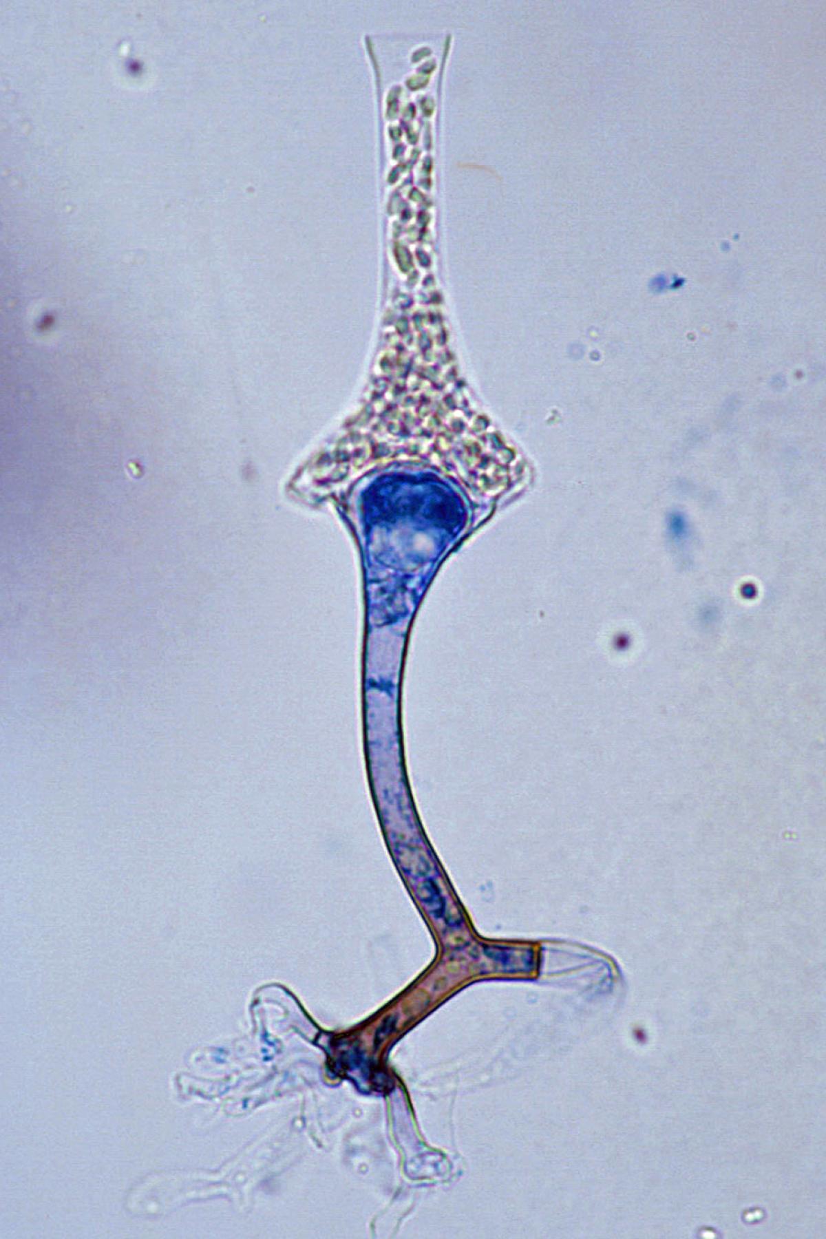
Sporangium of Saksenaea vasiformis.
Morphological description:
Colonies are fast growing, downy, white with no reverse pigment, and made up of broad, non-septate hyphae typical of a mucormycetous fungus. Sporangia are typically flask-shaped with a distinct spherical venter and long-neck, arising singly or in pairs from dichotomously branched, darkly pigmented rhizoids. Collumellae are prominent and dome-shaped. Sporangiospores are small, oblong, 1-2 x 3-4 µm, and are discharged through the neck following the dissolution of an apical mucilaginous plug.
Key features:
Mucorales, unique flask-shaped sporangia, failure to sporulate on primary isolation media.
Molecular identification:
ITS sequencing is required for differentiation of species within the complex, and may be necessary to achieve identification in a timely manner (Alvarez et al. 2010b, Walther et al. 2012, Halliday et al. 2015).
References:
Saksena (1953), Ellis and Hesseltine (1966), Ajello et al. (1976), Ellis and Ajello (1982), Ellis and Kaminski (1985), Pritchard et al. (1986), Padhye et al. (1988), Padhye and Ajello (1988), Goldschmied-Reouven et al. (1989), de Hoog et al. (2000) and Ellis (2005b).
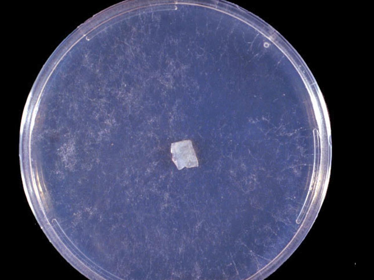
Culture of Saksenaea vasiformis.
Agar block method to induce sporulation of Saksenaea vasiformis. A small block of agar is cut from a well established culture grown on PDA and is placed in the centre of petri dish containing 1% agar in distilled water. After 21 days at 26C look for sporangium formation at the periphery of the petri dish.
Comment:
Laboratory identification of this fungus may be difficult or delayed because of the mould’s failure to sporulate on primary isolation media or on subsequent subculture onto potato dextrose agar. Sporulation may be stimulated by using the agar block method described by Ellis and Ajello (1982), Ellis and Kaminski (1985) and Padhye and Ajello (1988), although this may still take a period of days to weeks. Failure to sporulate prohibits antifungal susceptibility testing.
| Antifungal | Range | MIC90 | Antifungal | Range | MIC90 |
|---|---|---|---|---|---|
| AMB | 0.125-2 | 2 | VORI | 0.5-4 | 4 |
| POSA | 0.016-0.03 | 0.03 | POSA | 0.016-0.25 | 0.25 |
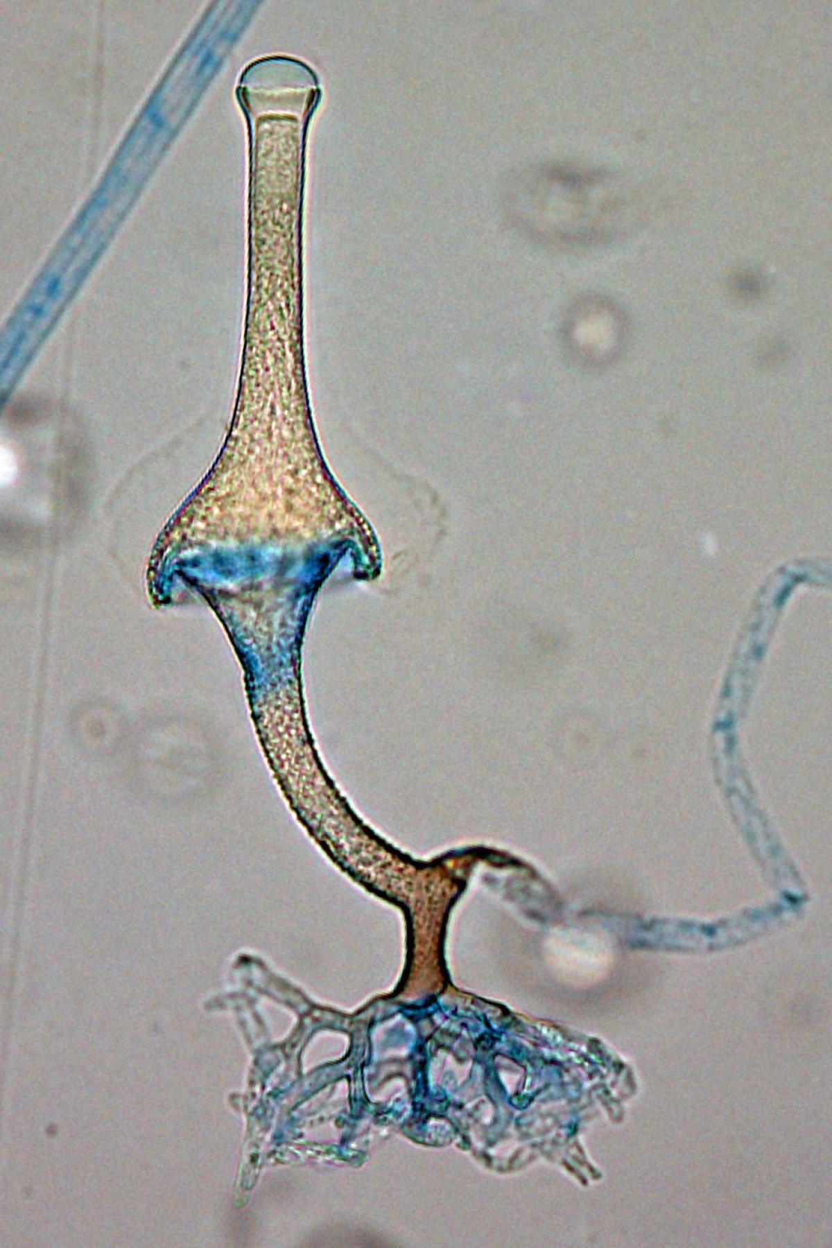
Saksenaea vasiformis sporangium showing distinctive rhizoids.