Status message
Correct! Excellent, you have really done well. Please find additional information below.
Unknown 62 = Exophiala spinifera
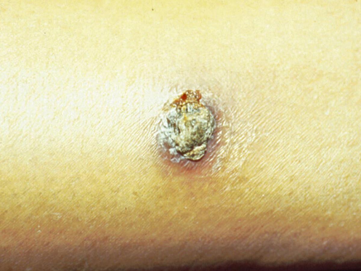
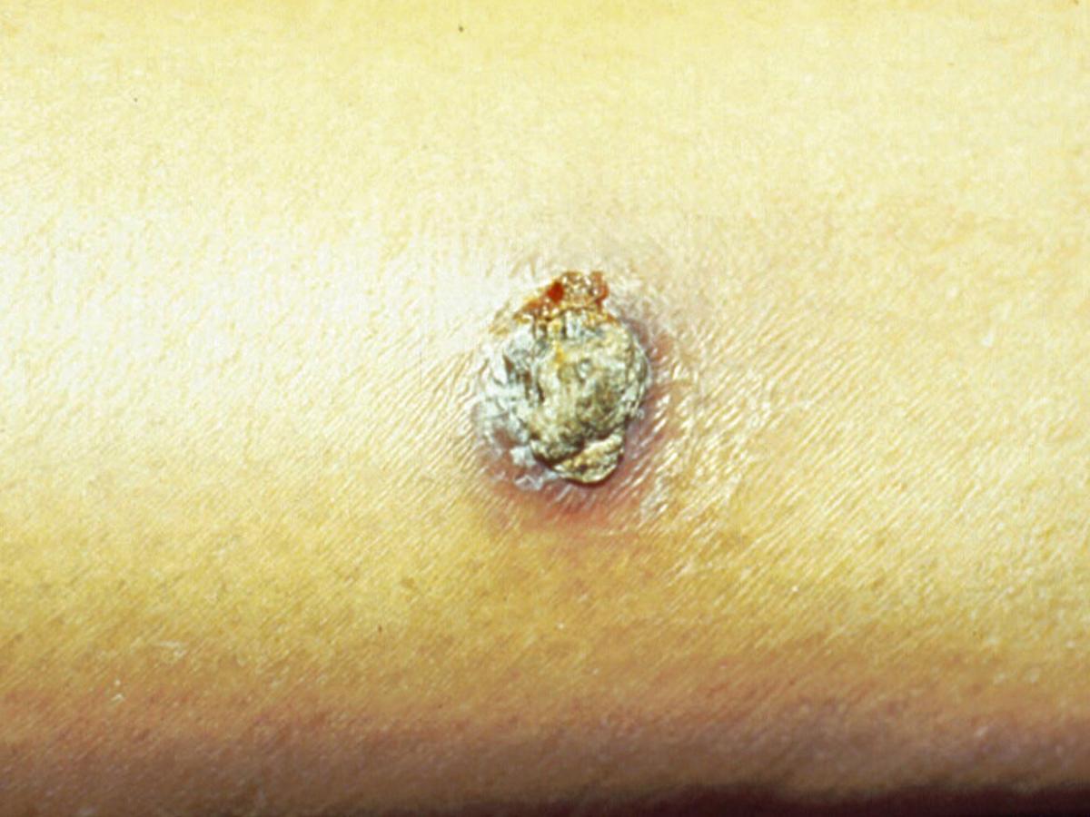
Clinical presentation: Cutaneous phaeohyphomycosis of the forearm caused by Exophiala spinifera.
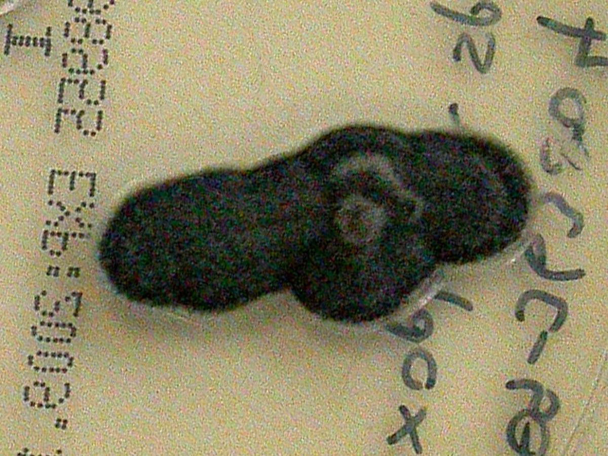
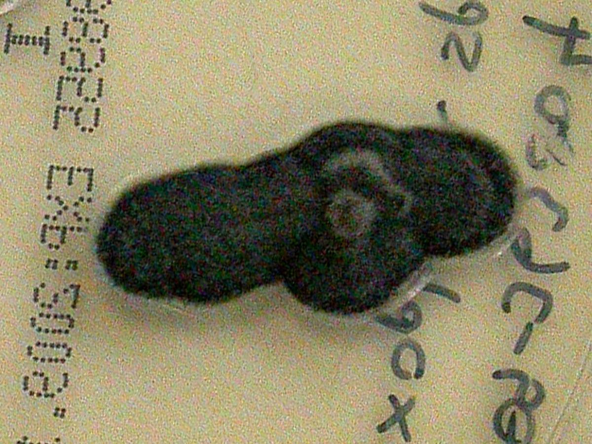
Culture: Colonies are initially mucoid and yeast-like, black, becoming raised and developing tufts of aerial mycelium with age, finally becoming suede-like to downy in texture. Reverse is olivaceous-black.
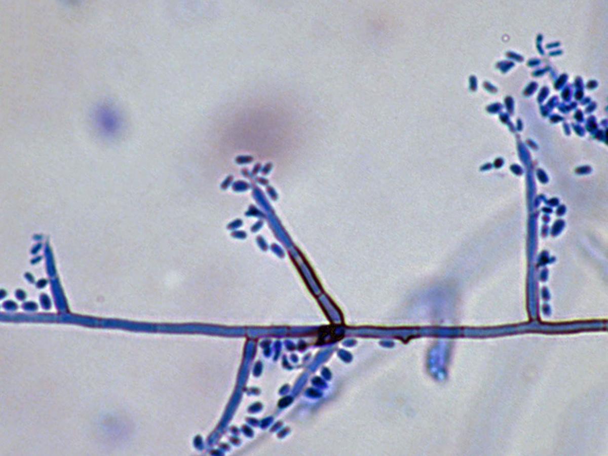
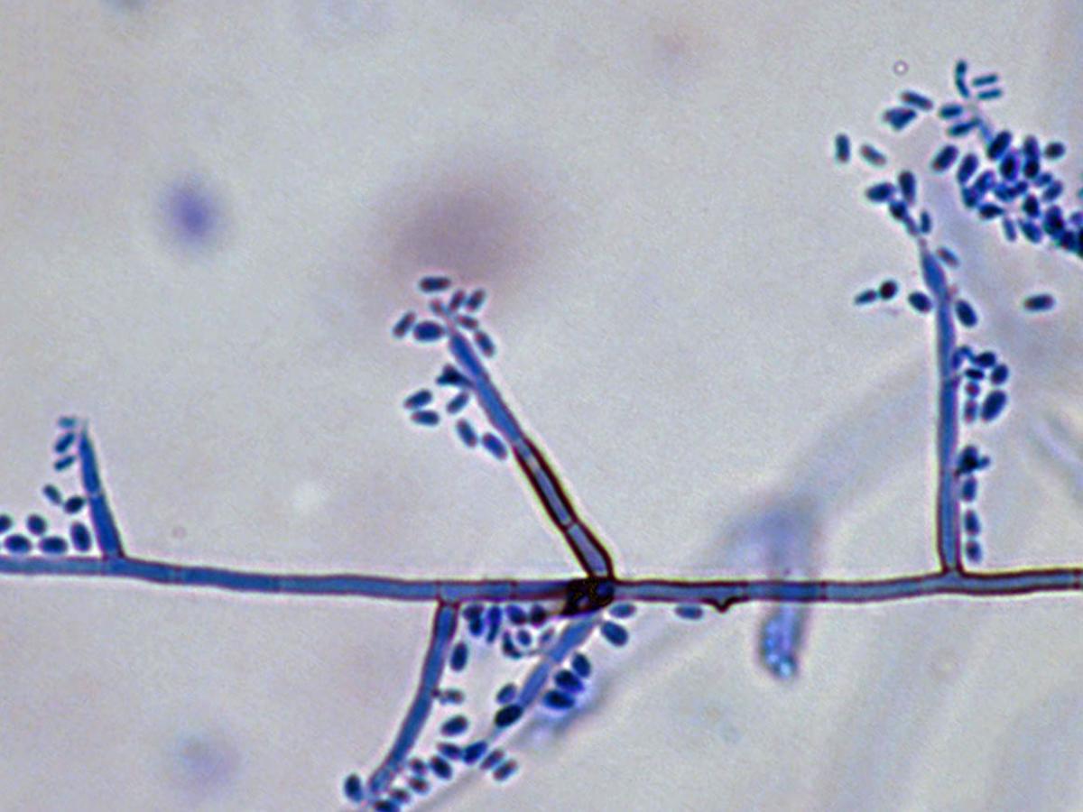
Microscopy: Conidiophores of Exophiala spinifera are simple or branched, erect or sub-erect, spine-like with rather thick brown pigmented walls. Conidia are formed on lateral pegs either arising apically or laterally at right or acute angles from the spine-like conidiophores or from undifferentiated hyphae. Conidia are one-celled, subhyaline, smooth, thin-walled, subglobose to ellipsoidal, and aggregate in clusters at the tip of each annellide. Toruloid hyphae and yeast-like cells with secondary conidia are typically present. No growth at 40C. RG-2 organism.
Comment: Recent molecular studies have re-examined Exophiala spinifera and have recognised two species: E. spinifera and E. attenuata (Vitale and de Hoog, 2002). These two species are morphologically very similar and can best be distinguished by genetic analysis. Conidiogenous cells are predominately annellidic and erect, multicellular conidiophores that are darker than the supporting hyphae are present. No growth at 40C.
| E. spinifera |
Annellated zones are long with clearly visible, frilled annellations. |
| E. attenuata |
Annellated zones are inconspicuous and degenerate. |
Exophiala species are common environmental fungi often associated with decaying wood and soil enriched with organic wastes. However, several species notably E. jeanselmei, E. moniliae and E. spinifera, are well documented human pathogens. Clinical manifestations include mycetoma (especially for E. jeanselmei), localized cutaneous infections, subcutaneous cysts, endocarditis and cerebral and disseminated infections. Phaeohyphomycosis caused by Exophiala species has been reported in both normal and immunosuppressed patients.
About Exophiala Back to virtual assessment
