Rhizopus
A molecular phylogenetic study of the genus Rhizopus by Abe et al. (2010) recognised eight species; R. caespitosus, R. delemar, R. homothallicus, R. microsporus, R. arrhizus (R. oryzae), R. reflexus, R. schipperae and R. stolonifer.
Based on these results and confirmed by Dolatabadi et al. (2014) the previous varieties of Rhizopus microsporus (R. microsporus var. oligosporus and R. microsporus var. rhizopodiformis) have been reduced to synonyms. In addition, R. azygosporus has been reduced to a synonym of R. microsporus. Finally, the controversy surrounding which species name to use for R. oryzae - R. arrhizus has been resolved in favour of the latter (Ellis 1985, de Hoog et al. 2015). Thus the important medical pathogens have now been reduced to just R. arrhizus and R. microsporus. These two species are the most common causative agents of mucormycosis, accounting for some 60% of the reported cases.
Morphological description:
The genus Rhizopus is characterised by the presence of stolons and pigmented rhizoids, the formation of sporangiophores, singly or in groups from nodes directly above the rhizoids, and apophysate, columellate, multispored, generally globose sporangia. After spore release the apophyses and columella often collapse to form an umbrella-like structure. Sporangiospores are globose to ovoid, one-celled, hyaline to brown and striate in many species. Colonies are fast growing and cover an agar surface with a dense cottony growth that is at first white becoming grey or yellowish brown with sporulation.
Molecular identification:
ITS sequencing is recommended but sequences must be compared to those of quality controlled reference strains with updated species names. (Alvarez et al. 2009 and Abe et al. 2010).
References:
Domsch et al. (1980), McGinnis (1980), Onions et al. (1981), Scholer et al. (1983), Schipper (1984), Schipper and Stalpers (1984, 2003), Yuan and Jong (1984), Ellis (1985, 1986), Rippon (1988), Kwon-Chung and Bennett (1992), Samson et al. (1995), Schipper et al. (1996), de Hoog et al. (2000, 2015), Ellis (2005b), Alvarez et al. (2009), Abe et al.(2010), Dolatabadi et al. (2014).
Species descriptions
-
Rhizopus arrhizus
Culture and sporangium with collapsed columella and sporangiospores of Rhizopus arrhizus.
Synonymy:
Rhizopus oryzaeRG-2 organism.
Morphological description:
Colonies are very fast growing, about 5-8 mm high, with some tendency to collapse, white cottony at first becoming brownish grey to blackish-grey depending on the amount of sporulation. Sporangiophores up to 1500 µm in length and 18 µm in width, smooth-walled, non-septate, simple or branched, arising from stolons opposite rhizoids usually in groups of three or more.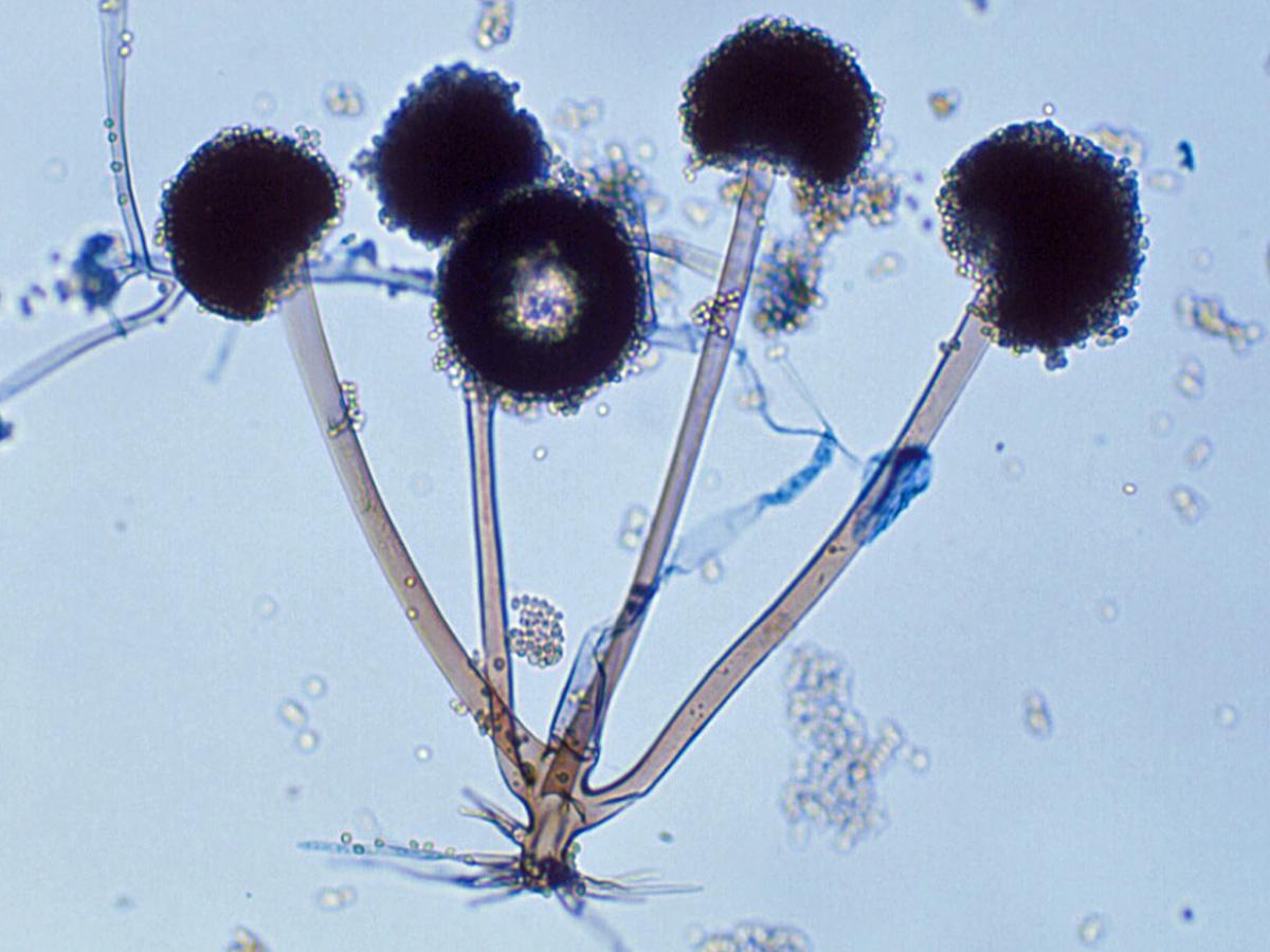
Sporangiophores, rhizoids and sporangia of Rhizopus arrhizus.
Sporangia are globose, often with a flattened base, greyish black, powdery in appearance, up to 175 µm in diameter and many spored. Columellae and apophysis together are globose, subglobose or oval, up to 130 µm in height collapsing to an umbrella-like form after spore release. Sporangiospores are angular, subglobose to ellipsoidal, with striations on the surface, and up to 8 µm in length. No growth at 45C; good growth at 40C.
Antifungal susceptibility: Rhizopus arrhizus (Australian national data); MIC µg/mL. Antifungal No <0.03 0.06 0.125 0.25 0.5 1 2 4 8 16 32 ≥64 AMB 53 4 12 20 14 2 1 ISAV 19 2 2 6 5 2 2 VORI 51 1 4 20 21 5 POSA 51 12 13 19 7 ITRA 53 1 4 12 18 6 2 2 8 -
Rhizopus microsporus
Synonymy:
Rhizopus azygosporus; Rhizopus microsporus var. microsporus; Rhizopus microsporus var. oligosporus; Rhizopus microsporus var. rhizopodiformis; Rhizopus microsporus var. chinensis.RG-2 organism.
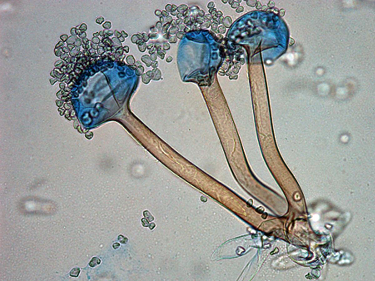
Rhizopus microsporus sporangia showing sporangiospores, columellae, sporangiophores and rhizoids.
Morphological description:
Colonies are dark greyish-brown, up to 10 mm high producing simple rhizoids. Sporangiophores are brownish, up to 400 µm high and 10 µm wide, and may be produced in groups of one to four, usually in pairs. Sporangia are greyish-black, spherical, up to 100 µm in diameter. Columellae are subglobose to globose to conical comprising 80% of the sporangium. Sporangiospores are angular to broadly ellipsoidal or subglobose, up to 5-9 µm in length and are distinctly striate. Chlamydospores may be present. Zygospores are dark red–brown, spherical, up to 100 µm in diameter, with stellate projections and unequal suspensor cells. Some strains may be homothallic and produce azygospores. There is good growth at 45C, with a maximum of 50-52C.Antifungal susceptibility: Rhizopus microsporus (Australian national data); MIC µg/mL. Antifungal No <0.03 0.06 0.125 0.25 0.5 1 2 4 8 16 32 ≥64 AMB 120 13 32 51 14 9 1 ISAV 23 2 7 10 3 1 VORI 120 3 27 87 3 POSA 120 3 10 55 35 16 1 ITRA 120 1 3 24 58 11 7 2 13 1