Status message
Correct! Excellent, you have really done well. Please find additional information below.
Unknown 61 = Microsporum canis
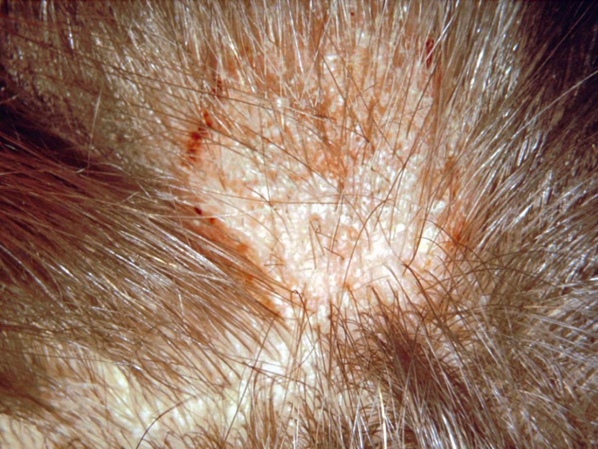
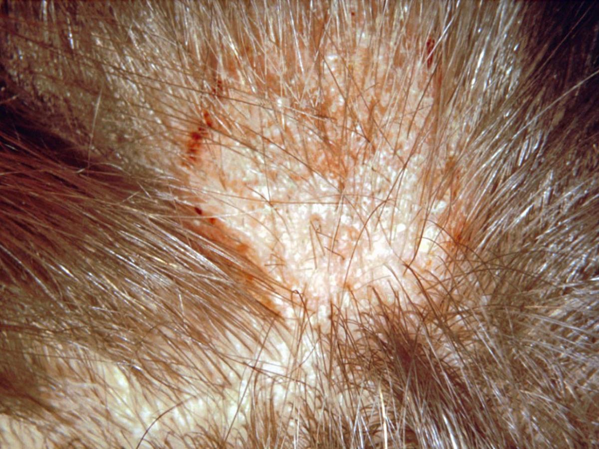
Clinical presentation: Early infection of the scalp by M. canis showing hair loss, scaling and advancing erythematous border.
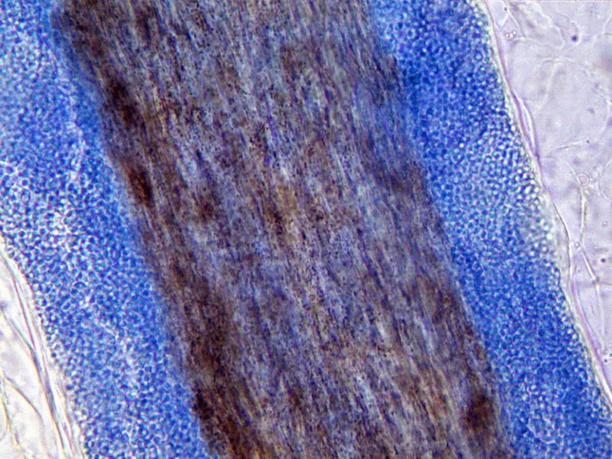
Direct microscopy (infected hair in KOH Parker ink preparation): Invaded hairs show an ectothrix infection and fluoresce a bright greenish-yellow under Wood's ultra-violet light.
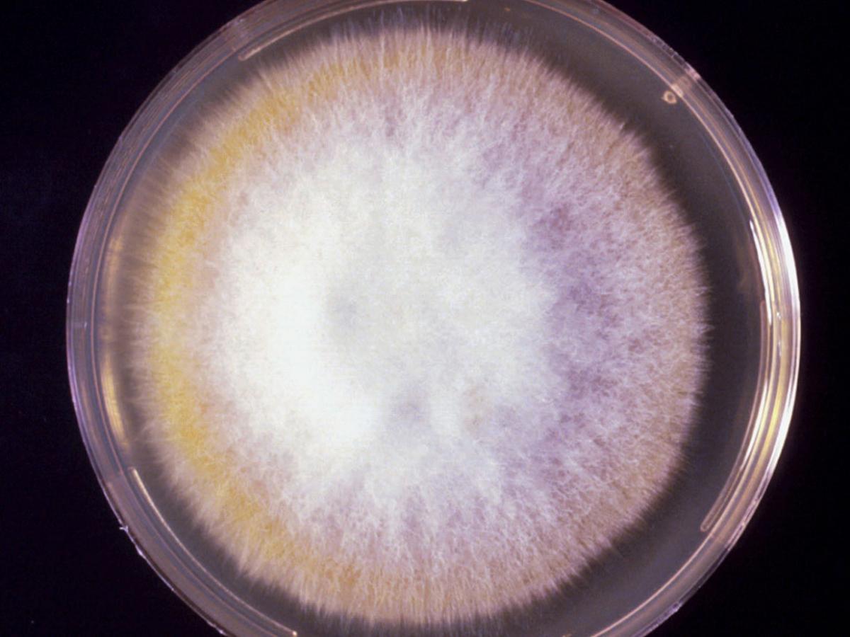
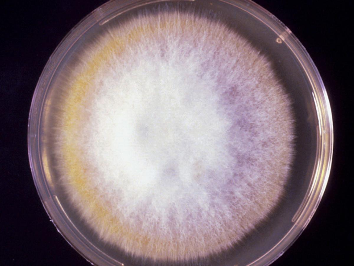
Culture: Colonies of M. canis are flat, spreading, white to cream-coloured, with a dense cottony surface which may show some radial grooves. Colonies usually have a bright golden yellow to brownish yellow reverse pigment, but non-pigmented strains may also occur.
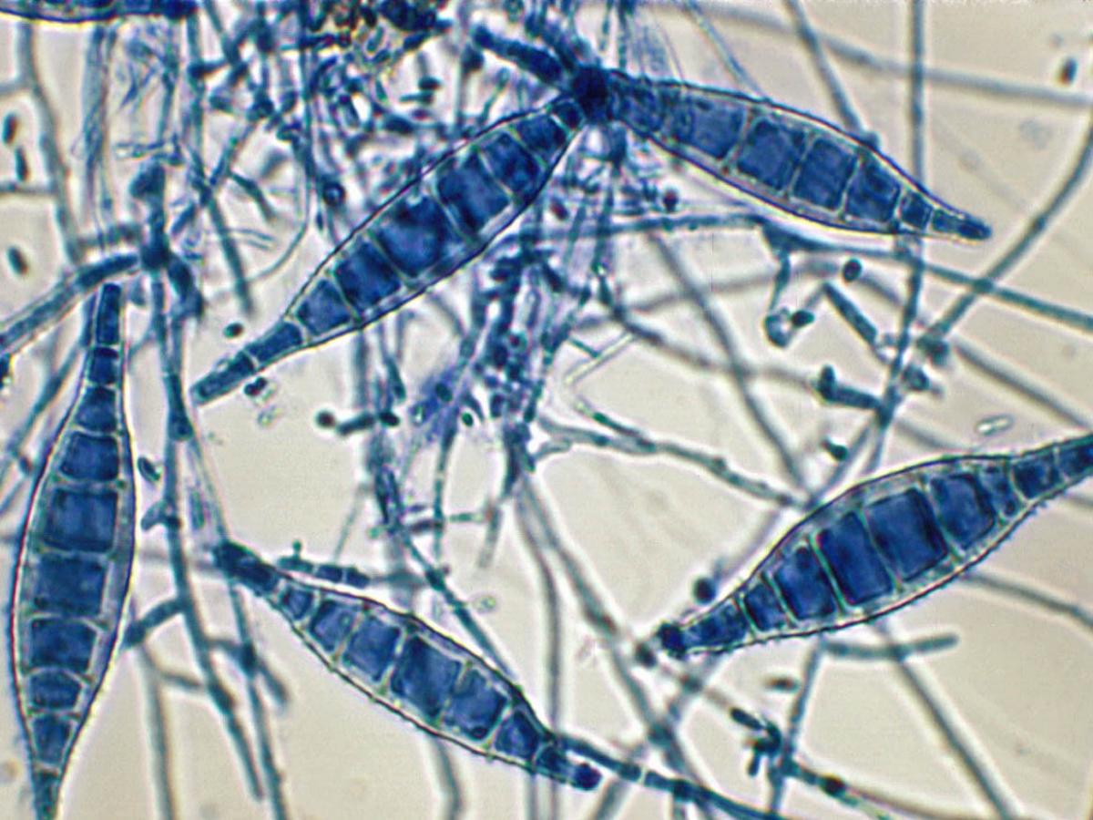
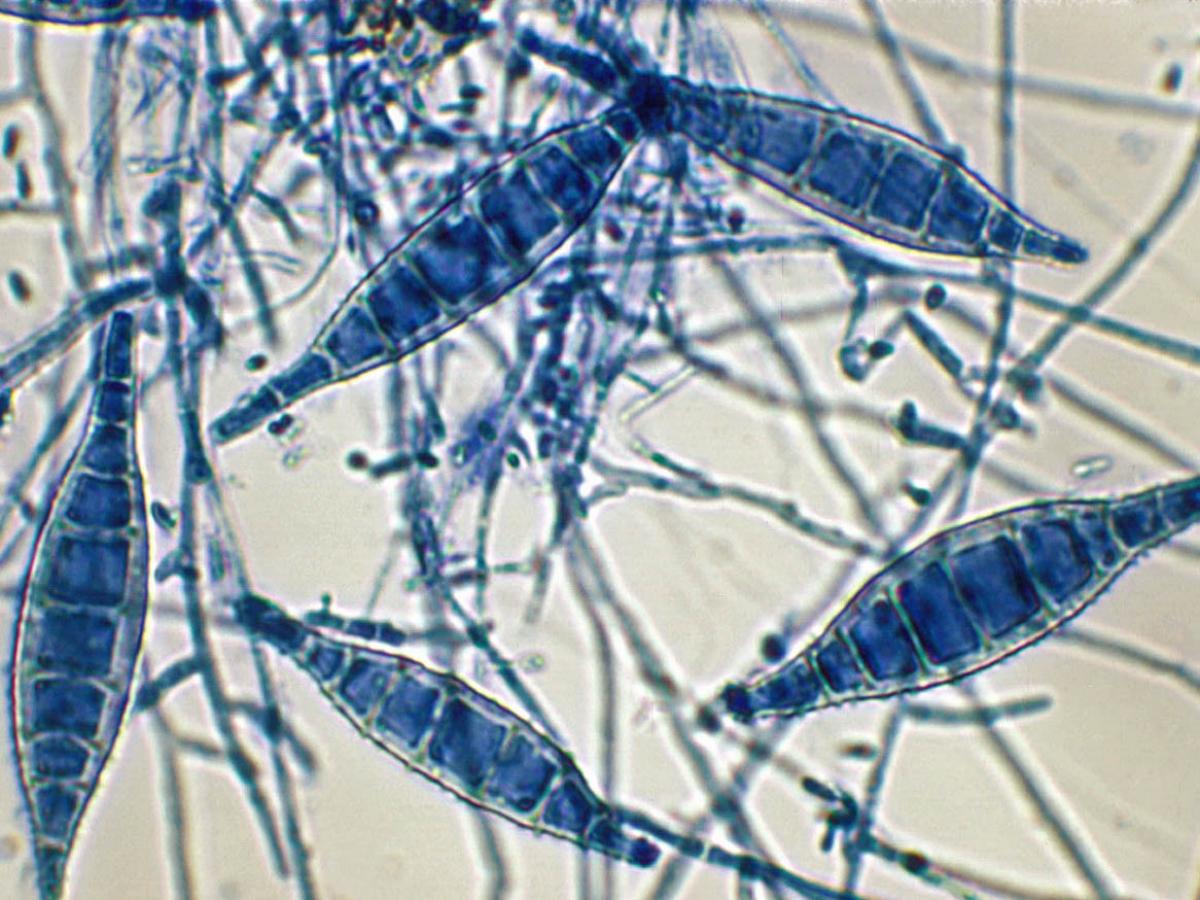
Microscopy: Macroconidia of M. canis are typically spindle-shaped with 5-15 cells, verrucose, thick-walled and often have a terminal knob, 35-110 x 12-25µm. A few pyriform to clavate microconidia are also present. Macroconidia and/or microconidia are often not produced on primary isolation media and it is recommended that sub-cultures be made onto Lactritmel Agar and/or boiled polished rice grains to stimulate sporulation. RG-2 organism.
Comment: Microsporum canis is a zoophilic dermatophyte of world-wide distribution which is a frequent cause of ringworm in humans, especially children. Invades hair, skin and rarely nails. Cats and dogs are the main sources of infection. Invaded hairs show an ectothrix infection and fluoresce a bright greenish-yellow under Wood's ultra-violet light.
About Microsporum Back to virtual assessment
