Status message
Correct! Excellent, you have really done well. Please find additional information below.
Unknown 74 = Phialophora verrucosa
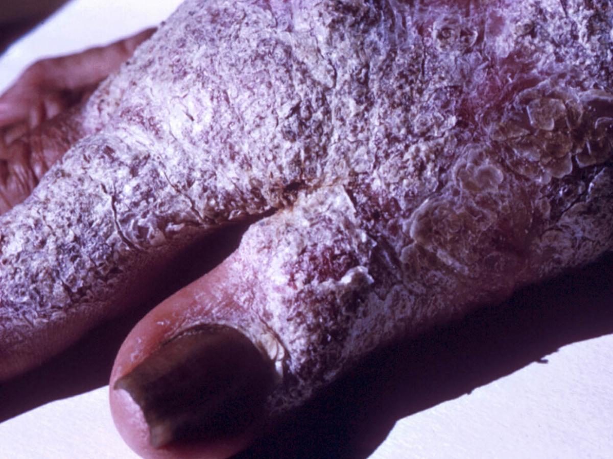
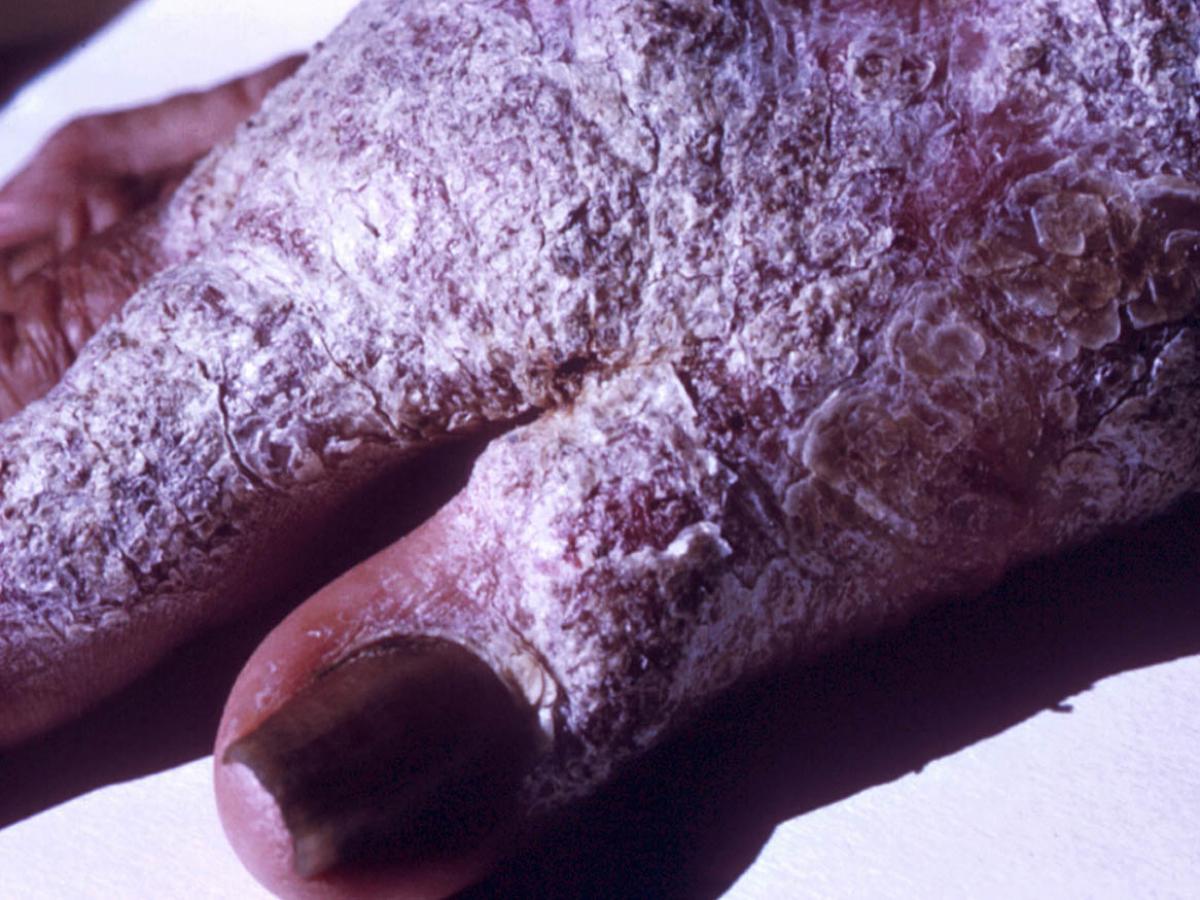
Clinical presentation: Chronic verrucous chromoblastomycosis of the hand. Note tissue hyperplasia forming a white verrucoid cutaneous lesion. In Australia, chromoblastomycosis occurs mostly on the hands and arms of timber and cattle workers in humid tropical forests.
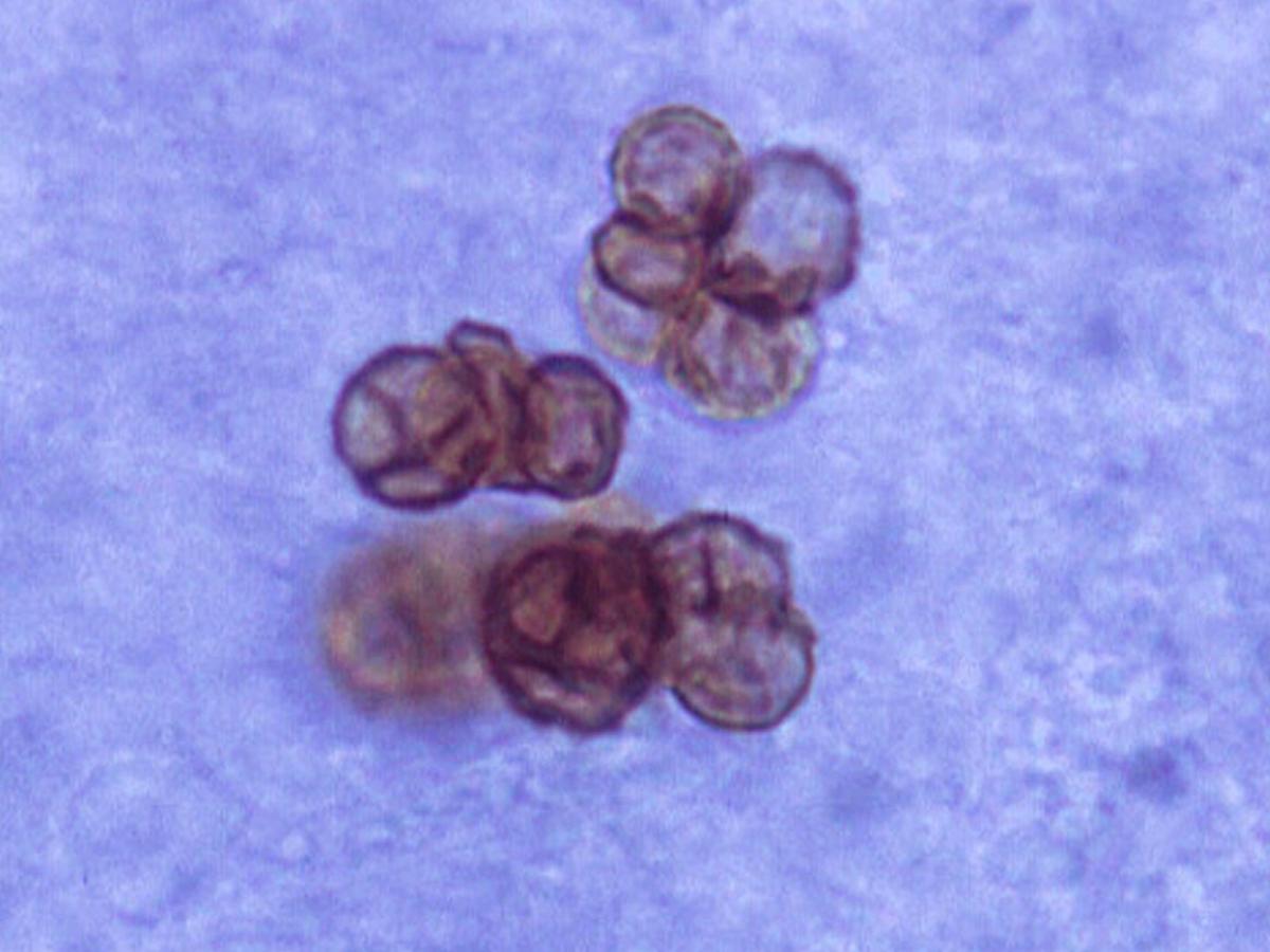
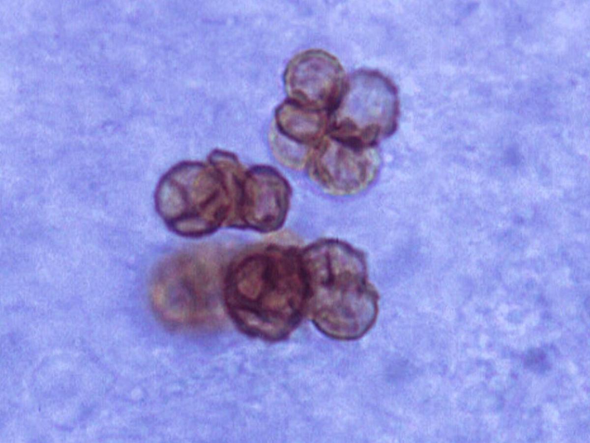
Direct microscopy: Skin scrapings from a patient with chromoblastomycosis mounted in 10% KOH and Parker ink solution showing characteristic brown pigmented, planate-dividing, rounded sclerotic bodies.
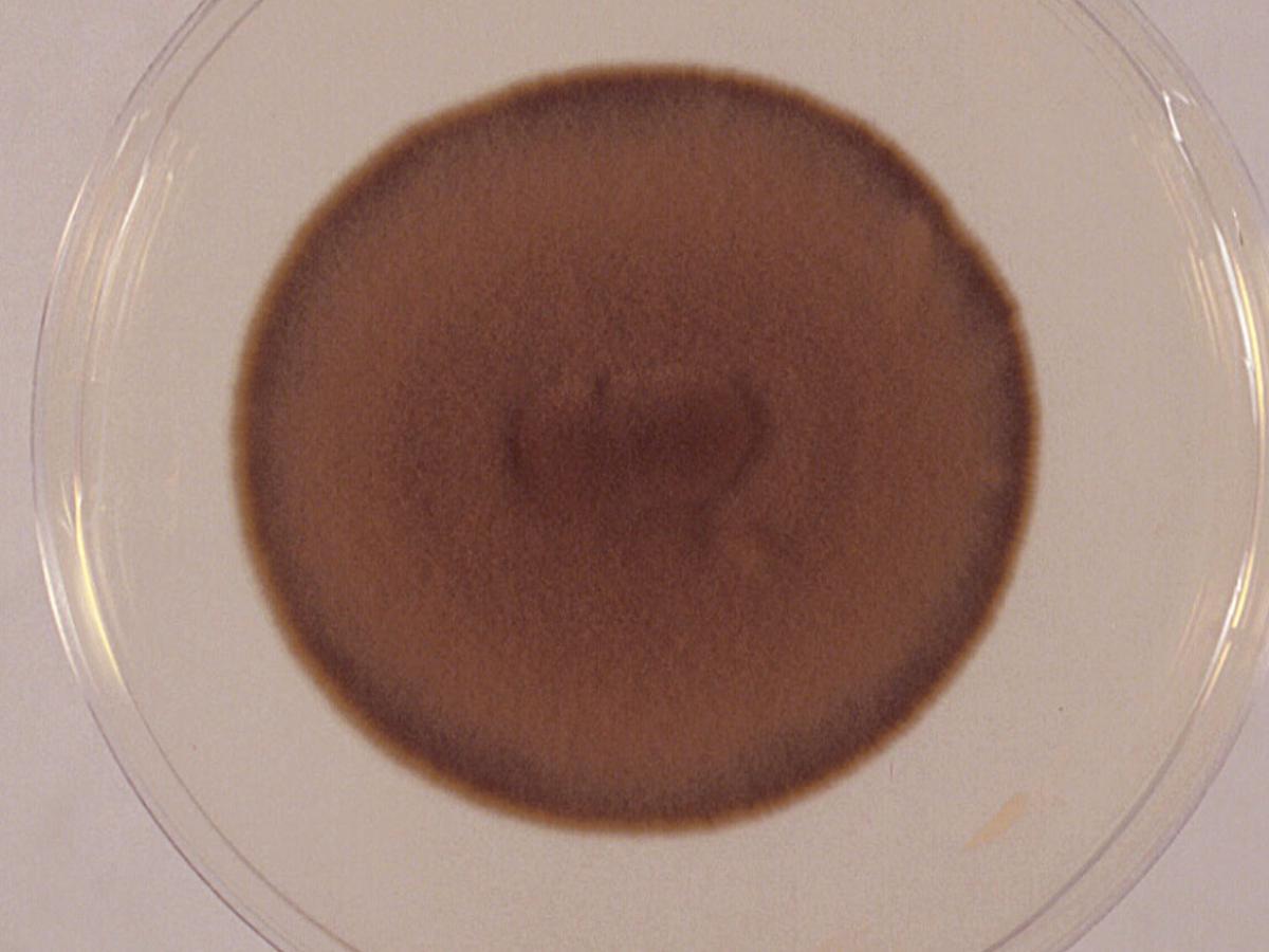
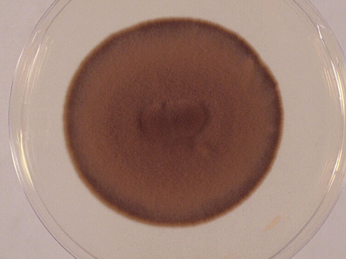
Culture: Colonies of Phialophora verrucosa are slow growing, initially dome-shaped, later becoming flat, suede-like and olivaceous to black in colour.
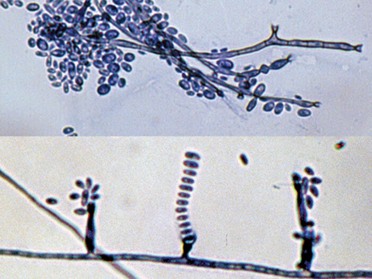
Microscopy: Phialides of Phialophora verrucosa are flask-shaped or elliptical with distinctive funnel-shaped, darkly pigmented collarettes. Conidia are ellipsoidal, smooth-walled, hyaline, mostly 3.0-5.0 x 1.5-3.0 μm, and aggregate in slimy heads at the apices of the phialide. RG-2 organism.
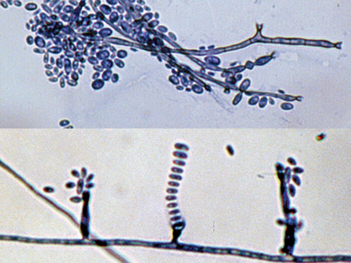
Comment: Phialophora verrucosa is a well documented causative agent of chromoblastomycosis, and mycetoma. It produces characteristic flask-shaped phialides with distinctive funnel-shaped, darkly pigmented collarettes. Environmental isolations have been made from plant debris, wood piles, fence posts, tree stumps, soil and animal faeces.
About Phialophora Back to virtual assessment
