Transmission Electron Microscopes (TEM)

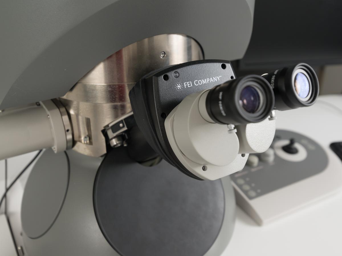
FEI Titan Themis 80-200
Please contact Dr Ashley Slattery for further information.
Located at Frome Road.
-
Information on the FEI Titan Themis 80-200
The FEI Titan Themis 80-200 is an ultra high resolution Transmission Electron Microscope enabling chemical and structural investigation of complex materials in the nano (billionths of a meter and less!) regime.
The Titan incorporates:
- Highest brightness X-FEG electron source
- Highly flexible accelerating voltage (80kV-200kV)
- Aberration correction on the probe forming lens (DCOR)
- High precision, Piezo driven specimen stage
- Latest technology in EDS detector technology (SuperX)
- Gatan Quantum GIF 965 Electron energy loss spectrometer
- Tomography specimen holder and software for 3D reconstructions
All this delivers a highly flexible and stable platform to routinely extract chemical and spatial information at the atomic level from complex materials.

FEI Tecnai G2 Spirit TEM
Please contact Mr Chris Leigh for further information.
Located at Frome Road.
-
Information on the FEI Tecnai G2 Spirit TEM
Operating at voltages of 20-120kV, the G2 Spirit is equipped with a FEG LaB6 emitter and BioTWIN lens design. This makes the G2 Spirit ideally suited to high contrast imaging of delicate light element biological materials typically found in life sciences. Imaging is done via an in-column Olympus-SIS Veleta CCD camera.
Installed options include cryo blades for cryo EM work, and capability for single and dual-axis 3D tomography using the Xplore 3D software. Analytical capabilities come via an EDS system comprising an Apollo XLT SDD running EDAX's TEAM software.

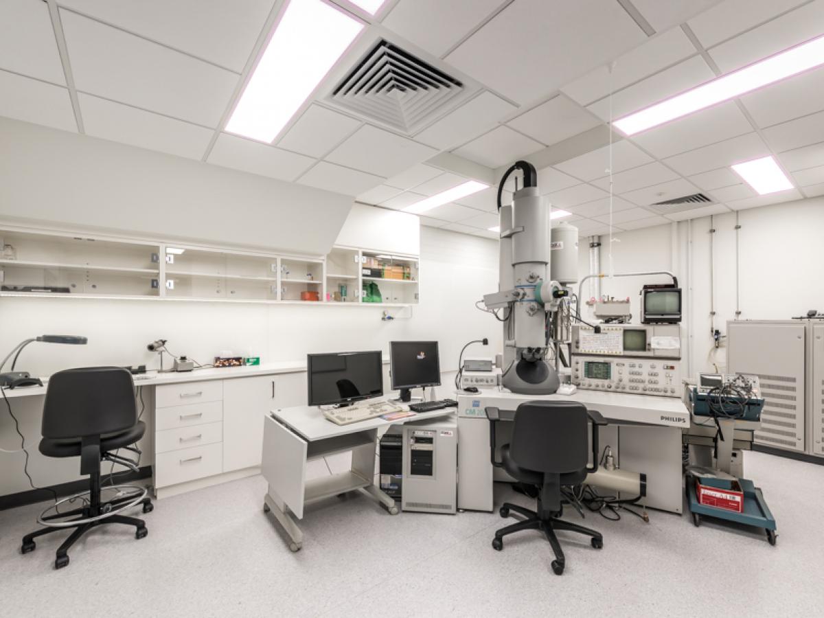
Philips CM200 Transmission Electron Microscope
Please contact Dr Ashley Slattery for further information.
Located at Frome Road.
-
Information on the Philips CM200 Transmission Electron Microscope
The Philips CM200 TEM is a high resolution Transmission Electron Microscope capable of operating up to 200kV.
Attachments include a full cryo setup with an EDAX EDS system and Gatan 678 Image Filter and P/EELS, and Gatan 832 SC1000 CCD camera, allowing for full imaging and analytical capability. This makes it ideal for high resolution imaging and analysis of nanotechnology samples including nanoparticles and nanotubes. It is also used routinely for imaging of biological and mineralogical samples.
A transmission electron microscope (TEM) is comparable in design to an inverted light microscope. An electron gun at the top of the column produces a beam of electrons, which are accelerated down the column, and focused into a coherent beam by electromagnetic lenses onto the sample. The specimen, mounted on a small copper grid, must be thin enough to allow the beam to pass through it. The objective lens forms an image of the sample, which is further magnified by the imaging lenses below. The image is projected onto the fluorescent screen at the bottom of the column. In various positions in the column are a series of apertures, which serve to eliminate any stray electrons from the image forming process.
RECOMMENDED SAMPLE SIZE: Grid size is 3mm diameter, samples should be <100nm thick. Initial sample fixation for biological samples should not exceed 1mm³ (prior to sample dehydration & embedding). Contact us for protocols.
RESOLUTION: 0.2nm
MAGNIFICATION: 2,000,000x
The interaction of the electron beam with the sample also generates other signals (e.g. X-rays) which can be used on the CM200 to give further information about the specimen using EDAX. X-rays generated by the interaction of the electron beam and sample are characteristic of the elements in the sample. The above example (Label A) is a spectrum obtained from the multi-layer coating on an ophthalmic lens, and shows the presence of silicon (Si) and oxygen (O) in the outer layer. The technique is non-destructive and is quantitative over the elemental range boron (B, Z=5) to uranium (U, Z=92). Software packages are available for particulate sizing and characterisation, and for elemental distribution maps.

Philips CM100 Transmission Electron Microscope
Please contact Dr Gwen Mayo for further information.
Located at Waite facility.
-
Information on the Philips CM100 Transmission Electron Microscope
The Philips CM100 Transmission Electron Microscope has a maximum accelerating voltage of 100kV. It is equipped with a SIS MegaviewII CCD Camera and analySIS image analysis software.
This TEM is used primarily for biological samples and some materials samples when highest resolution is not required.
A transmission electron microscope (TEM) is comparable in design to an inverted light microscope. An electron gun at the top of the column produces a beam of electrons, which are accelerated down the column, and focused into a coherent beam by electromagnetic lenses onto the sample. The specimen, mounted on a small copper grid, must be thin enough to allow the beam to pass through it. The objective lens forms an image of the sample, which is further magnified by the imaging lenses below.
The image is projected onto the fluorescent screen at the bottom of the column. In various positions in the column are a series of apertures, which serve to eliminate any stray electrons from the image forming process.
RECOMMENDED SAMPLE SIZE: Grid size is 3mm diameter, samples should be <100nm thick. Initial sample fixation for biological samples should not exceed 1mm³ (prior to sample dehydration & embedding). Contact us for protocols.
RESOLUTION: 0.24nm
MAGNIFICATION: 100,000x

FEI Glacios 200kV Cryo-Transmission Electron Microscope
Please contact Dr Fiona Whelan for further information.
Located at Frome Road.
-
Information on the FEI Glacios 200kV Cryo-Transmission Electron Microscope
The Thermo Scientific Glacios Cryo-Transmission Electron Microscope (Cryo-TEM) delivers a complete cryo-electron microscopy (cryo-EM) solution to a broad range of scientists. It features 200 kV XFEG optics, the industry-leading Autoloader (cryogenic sample manipulation robot), and the same innovative automation for ease of use as on the Thermo Scientific Krios G4 Cryo-TEM. The Glacios Cryo-TEM bundles all this into a smaller footprint that simplifies installation.
The designed-in connectivity ensures a robust and contamination-free pathway throughout the entire workflow, from sample preparation and optimization to image acquisition and data processing. The Glacios Cryo-TEM can be used in a single particle analysis workflow for pre-optimisation of sample quality, as well as for high-resolution single particle analysis data acquisition. Furthermore, the Glacios Cryo-TEM can also be configured with a direct electron detector and/or Volta phase plate to be a complete standalone single particle analysis data acquisition solution. Pairing the Glacios Cryo-TEM with the Thermo Scientific Selectris and Selectris X Imaging Filters can be used as a complete solution for single particle analysis data acquisition and cryo-electron tomography (cryo-ET). Overall, the Glacios Cryo-TEM is a versatile microscope covering all ranges of applications such as single particle analysis, cryo-ET, as well microcrystal electron diffraction (MicroED).
